. 2016 Jul 1;30(13):1542-57.
doi: 10.1101/gad.284166.116.

HGF-independent regulation of MET and GAB1 by nonreceptor tyrosine kinase FER potentiates metastasis in ovarian cancer

Affiliations

HGF-independent regulation of MET and GAB1 by nonreceptor tyrosine kinase FER potentiates metastasis in ovarian cancer

Gaofeng Fan et al. Genes Dev. .

Abstract

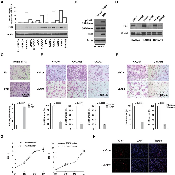
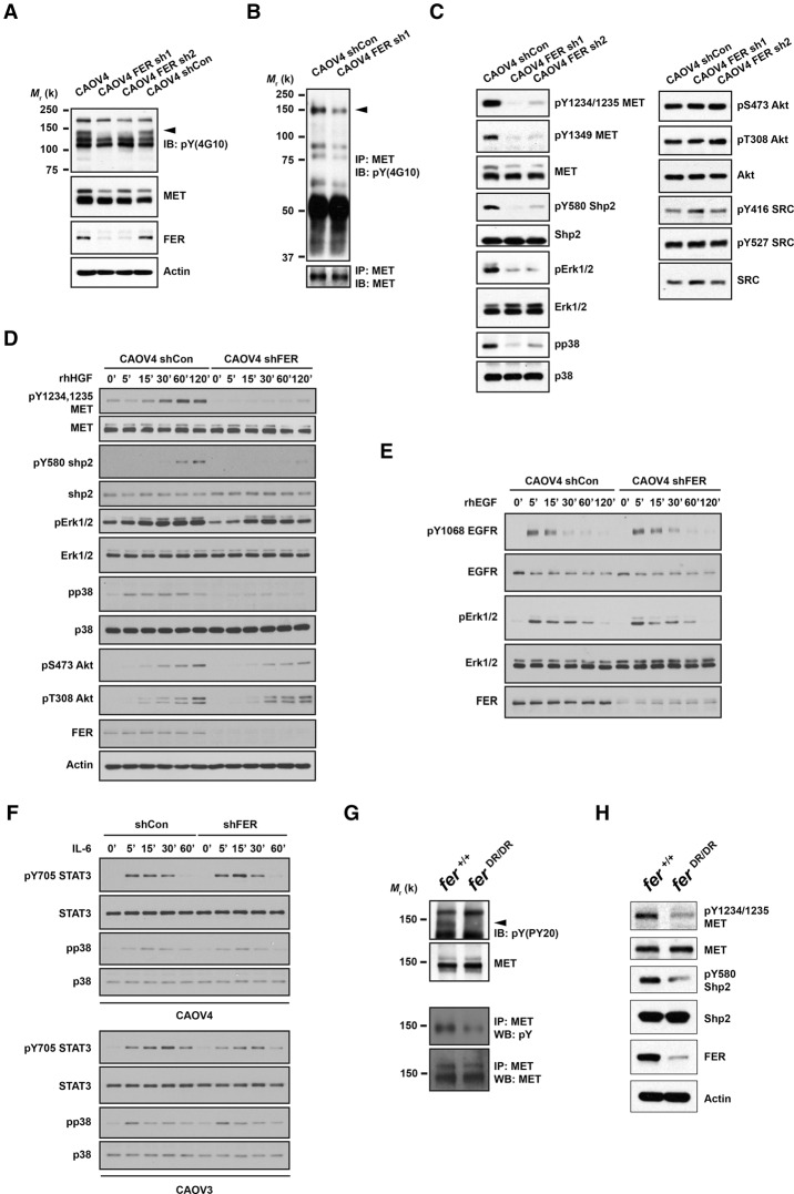
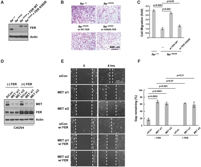
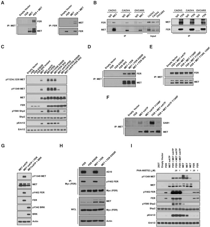
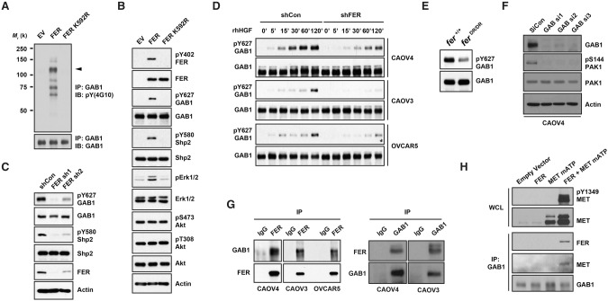
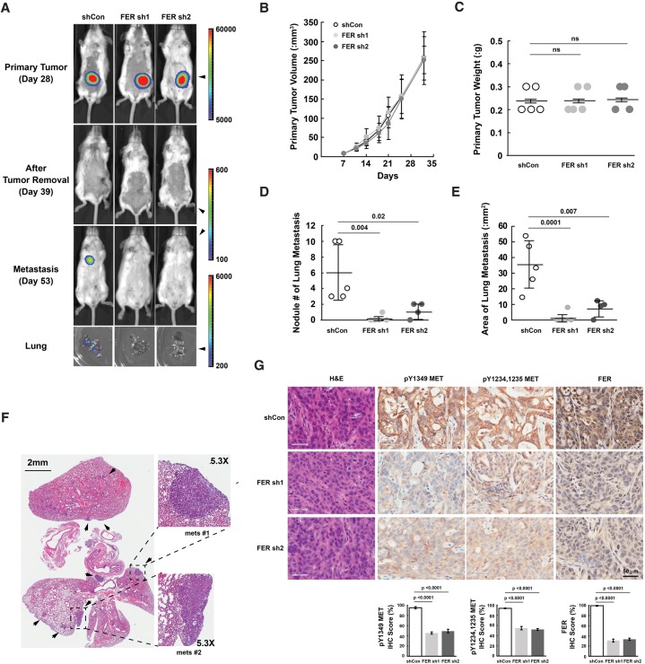
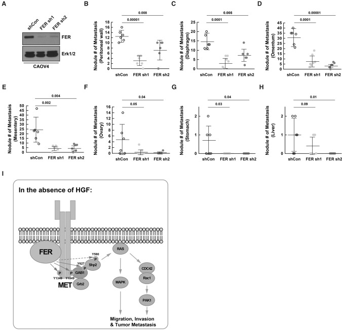
Ovarian cancer cells disseminate readily within the peritoneal cavity, which promotes metastasis, and are often resistant to chemotherapy. Ovarian cancer patients tend to present with advanced disease, which also limits treatment options; consequently, new therapies are required. The oncoprotein tyrosine kinase MET, which is the receptor for hepatocyte growth factor (HGF), has been implicated in ovarian tumorigenesis and has been the subject of extensive drug development efforts. Here, we report a novel ligand- and autophosphorylation-independent activation of MET through the nonreceptor tyrosine kinase feline sarcoma-related (FER). We demonstrated that the levels of FER were elevated in ovarian cancer cell lines relative to those in immortalized normal surface epithelial cells and that suppression of FER attenuated the motility and invasive properties of these cancer cells. Furthermore, loss of FER impaired the metastasis of ovarian cancer cells in vivo. Mechanistically, we demonstrated that FER phosphorylated a signaling site in MET: Tyr1349. This enhanced activation of RAC1/PAK1 and promoted a kinase-independent scaffolding function that led to recruitment and phosphorylation of GAB1 and the specific activation of the SHP2-ERK signaling pathway. Overall, this analysis provides new insights into signaling events that underlie metastasis in ovarian cancer cells, consistent with a prometastatic role of FER and highlighting its potential as a novel therapeutic target for metastatic ovarian cancer.

Keywords: FER; GAB1; MET; ovarian cancer; tyrosine phosphorylation.


Figures

Figure 1.
Figure 1.
FER was up-regulated in ovarian cancer-derived cell lines and was essential for cell motility and invasiveness. (A) Total cell lysates from two control and 11 ovarian carcinoma-derived cell lines were immunoblotted for FER and loading control actin. (B) HOSE11–12 cells were transiently transfected with either empty vector or FER constructs and immunoblotted for tyrosine phosphorylation of β-catenin, FER, and a loading control, ERK. (C) The effect of FER overexpression on cell motility was measured by Boyden chamber assay 24 h after seeding. Representative bright-field images are illustrated, together with quantitation. Mean ± S.E.M. n = 3. (D) shRNA knockdown of FER in three ovarian cancer-derived cell lines. (E,F) The effects of FER loss on cell motility (E) and invasiveness (F) were measured by Boyden chamber assay 24 h after seeding. Representative bright-field images are illustrated, together with quantitation. Mean ± S.E.M. n = 3. (G,H) Cell growth was assessed by CellTiter-Glo luminescent cell viability assay at the indicated time intervals (G) and by Ki-67 immunostaining (H).
Figure 2.
Figure 2.
Loss of FER attenuated tyrosine phosphorylation of HGFR (MET) and MET-mediated downstream signaling. (A) Anti-phosphotyrosine antibody immunoblot (4G10) to examine the impact of FER knockdown on tyrosine phosphorylation in CAOV4 cells. The blot was reprobed with antibodies against MET, FER, and the loading control actin. (B) Tyrosine phosphorylation of MET was examined by immunoprecipitation followed by blotting with anti-phosphotyrosine antibody 4G10. The blot was reprobed for MET to indicate equal immunoprecipitation efficiency and loading. (C) CAOV4 cells expressing either control or FER shRNA were lysed and immunoblotted as indicated to measure the activation of MET and MET-regulated downstream signaling pathways. (DF) Cells were serum-starved and stimulated with recombinant human HGF (D), EGF (E), or IL-6 (F) for the indicated times; lysed; and immunoblotted with the designated antibodies to illustrate the impact of FER deficiency on HGF-, EGF-, or IL-6-induced signaling. (G, top) Anti-phosphotyrosine antibody immunoblot (PY20) to examine the effects of a destabilized FER-D743R mutant on tyrosine phosphorylation in mouse embryonic fibroblasts (MEFs). The position of MET is indicated by an arrowhead. The blot was reprobed for MET. (Bottom) MET was immunoprecipitated from both Fer+/+ and FerDR/DR MEF cell lysates, and tyrosine phosphorylation was examined by immunoblotting with antibody 4G10. (H) Fer+/+ and FerDR/DR MEF cells were lysed and immunoblotted as indicated to illustrate the impact of FER destabilization on MET and MET-mediated downstream signaling pathways.
Figure 3.
Figure 3.
The kinase activity of FER and its downstream effector, MET, were essential for cell motility. (A) MEF cell lysates Fer+/+, FerDR/DR, and FerDR/DR rescued with either 6xMYC-tagged wild type or the kinase-dead FER-K592R mutant were immunoblotted with antibody against FER to demonstrate comparable expression. Actin was probed as the loading control. (B,C) A Boyden chamber assay was performed on the indicated MEF cells, in which migration was monitored 24 h after seeding. Representative images are illustrated (B), along with quantitation (mean ± SEM; n = 3) (C). (D) Both MET siRNAs and a FER expression construct were delivered as indicated into CAOV4 cells by electroporation. After 48 h, the expression levels of MET and FER were measured by immunoblotting, with actin as a loading control. (E,F) Forty-eight hours after electroporation and seeding, a wound healing assay was performed on the cell lines indicated in D. Wound recovery was recorded 6 h after scratch injury. Representative images are illustrated (E), together with quantitation (mean ± SEM; n = 3) (F).
Figure 4.
Figure 4.
FER bound to and phosphorylated MET at Tyr1349. (A) After transient transfection in 293T cells, MET was immunoprecipitated and probed for association with FER (left) and vice versa (right). (B) Endogenous MET was immunoprecipitated from CAOV4, CAOV3, and OVCAR5 ovarian cancer cells and probed for association with FER (left) and vice versa (right). (C) 293T cells were transiently transfected with the indicated constructs, lysed, and immunoblotted as indicated to illustrate the phosphorylation of MET by FER and its impact on downstream SHP2–ERK signaling. (D) MET was coexpressed with either wild-type or mutant forms of FER (kinase-dead K592R or SH2 mutant) in 293T cells to compare the effects of mutations in FER on its association with MET. (E) FER was coexpressed with either wild-type or Tyr→Phe mutants of MET (Y1349F, Y1356F, or YY1349,1356FF) in 293T cells to compare the effects of the MET point mutations on its association with FER. (F) FER was expressed alone or with either mATP or mATP Y1349F mutant forms of MET in 293T cells. MET was immunoprecipitated, and the association with GAB1 in each sample was assessed by immunoblotting. (G) 293T cells were transiently transfected with MET mATP alone or together with either FER or BRK, and the phosphorylation of MET on Tyr1349 was assessed by immunoblotting. (H) 6xMYC-tagged wild-type or inactive K592R FER was expressed alone or cotransfected with MET, immunoprecipitated with anti-MYC antibody, and probed for tyrosine phosphorylation with either 4G10 (global Tyr phosphorylation) or pTyr402-FER-specific antibodies. (I) Transfected 293T cells were treated with or without MET inhibitor PHA-665752 (4 h), as indicated, and the cell lysates were immunoblotted with the designated antibodies to illustrate the effect of the small molecule inhibitor on MET phosphorylation and downstream SHP2–ERK signaling.
Figure 5.
Figure 5.
FER formed a complex with GAB1 via MET and phosphorylated GAB1 at Tyr627. 293T cells were transiently transfected with either wild-type FER or a kinase-dead FER-K592R mutant. (A) Tyrosine phosphorylation of GAB1 was examined by immunoprecipitation and blotting with anti-phosphotyrosine antibody 4G10. (B) The whole-cell lysates were immunoblotted as indicated to show the increased tyrosine phosphorylation of GAB1 and SHP2 and the impact on downstream signaling. (C) Tyrosine phosphorylation of GAB1 and SHP2 was compared in CAOV4 cells expressing either control or FER targeted shRNAs. (D) Cells were serum-starved and stimulated with hHGF for the indicated times, lysed, and immunoblotted with both pTyr627 and total GAB1 antibodies to demonstrate FER-regulated GAB1 phosphorylation. (E) Phosphorylation status of Tyr627 of GAB1 in Fer+/+ and FerDR/DR MEF cells. (F) Control or GAB1 siRNAs were delivered into CAOV4 cells by electroporation. After 48 h, the expression levels of GAB1 and activation of PAK1 were measured by immunoblotting. (G) Endogenous FER was immunoprecipitated from CAOV4, CAOV3, and OVCAR5 ovarian cancer cells, and its association with GAB1 was examined by immunoblotting (left) and vice versa (right). (H) Endogenous GAB1 was immunoprecipitated from lysates of 293T cells transfected with FER and the MET mATP mutant alone or together. The association of FER and MET was assessed by immunoblotting.
Figure 6.
Figure 6.
Loss of FER reduced lung metastasis burden of ovarian cancer cell xenografts with inactivation of MET. (A) At the indicated time points, mice injected with CAOV4 cells expressing either shCon (n = 5), FER sh1 (n = 5), or FER sh2 (n = 4) were imaged using IVIS bioluminescence imaging. Representative images are shown. (B,C) Measurements of subcutaneous tumor volume (B) and weight (C) for mice injected with either shCon, FER sh1, or FER sh2 CAOV4 cells. (D) Metastasis lesion count (naked eye as well as IVIS imaging confirmation) with mice injected with either shCon, FER sh1, or FER sh2 CAOV4 cells. (E) Metastasis lesion area measurement with mice injected with either shCon, FER sh1, or FER sh2 CAOV4 cells. (F) Representative H&E staining of lung sections from mice with metastatic lesions that express the control shRNA (shCon). (G) H&E and immunohistochemistry staining (pTyr MET and FER) of subcutaneous tumor sections from mice injected with CAOV4 cells expressing either shCon, FER sh1, or FER sh2. The immunohistochemistry image was scored with Aperio software.
Figure 7.
Figure 7.
Loss of FER reduced metastasis of ovarian cancer cells via the peritoneal cavity. (A) Knockdown efficiency was confirmed by immunoblotting in both FER shRNA CAOV4 cell lines prior to intraperitoneal injection. (BH) Mice were injected intraperitoneally with 5 × 10⁶ CAOV4 cells expressing either shCon (n = 6), FER sh1 (n = 7), or FER sh2 (n = 6). After 4 wk, necropsy procedures were performed, and the ability of CAOV4 cells to metastasize to surrounding tissue/organs, including the peritoneal wall (B), diaphragm (C), omentum (D), mesentery (E), ovary (F), stomach (G), and liver (H), were assessed. (I) Working model: In the absence of the ligand HGF, FER directly phosphorylated MET, GAB1, and possibly SHP2. This led to the activation of SHP2–MAPK and RAC1–PAK1 signaling downstream from MET to potentiate the motility and invasiveness of ovarian cancer cells.

Similar articles

Cited by

References

    1. Ahn J, Truesdell P, Meens J, Kadish C, Yang X, Boag AH, Craig AW. 2013. Fer protein-tyrosine kinase promotes lung adenocarcinoma cell invasion and tumor metastasis. Mol Cancer Res 11: 952–963. - PubMed
    1. Albeck JG, Brugge JS. 2011. Uncovering a tumor suppressor for triple-negative breast cancers. Cell 144: 638–640. - PubMed
    1. Arend RC, Londono-Joshi AI, Straughn JM Jr, Buchsbaum DJ. 2013. The Wnt/β-catenin pathway in ovarian cancer: a review. Gynecol Oncol 131: 772–779. - PubMed
    1. Ayhan A, Ertunc D, Tok EC, Ayhan A. 2005. Expression of the c-Met in advanced epithelial ovarian cancer and its prognostic significance. Int J Gynecol Cancer 15: 618–623. - PubMed
    1. Barbolina MV, Burkhalter RJ, Stack MS. 2011. Diverse mechanisms for activation of Wnt signalling in the ovarian tumour microenvironment. Biochem J 437: 1–12. - PMC - PubMed

MeSH terms

Substances