. 2016 Jul 1;30(13):1558-72.
doi: 10.1101/gad.280222.116.

The Nrd1-like protein Seb1 coordinates cotranscriptional 3' end processing and polyadenylation site selection

Affiliations

The Nrd1-like protein Seb1 coordinates cotranscriptional 3' end processing and polyadenylation site selection

Jean-François Lemay et al. Genes Dev. .

Abstract

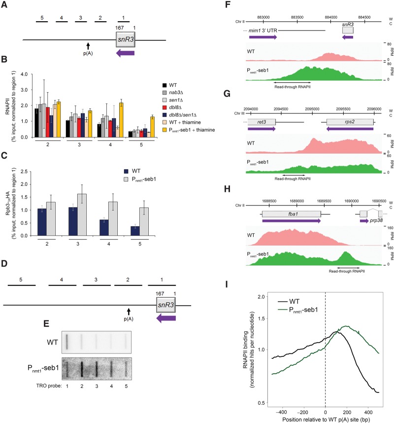
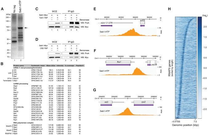
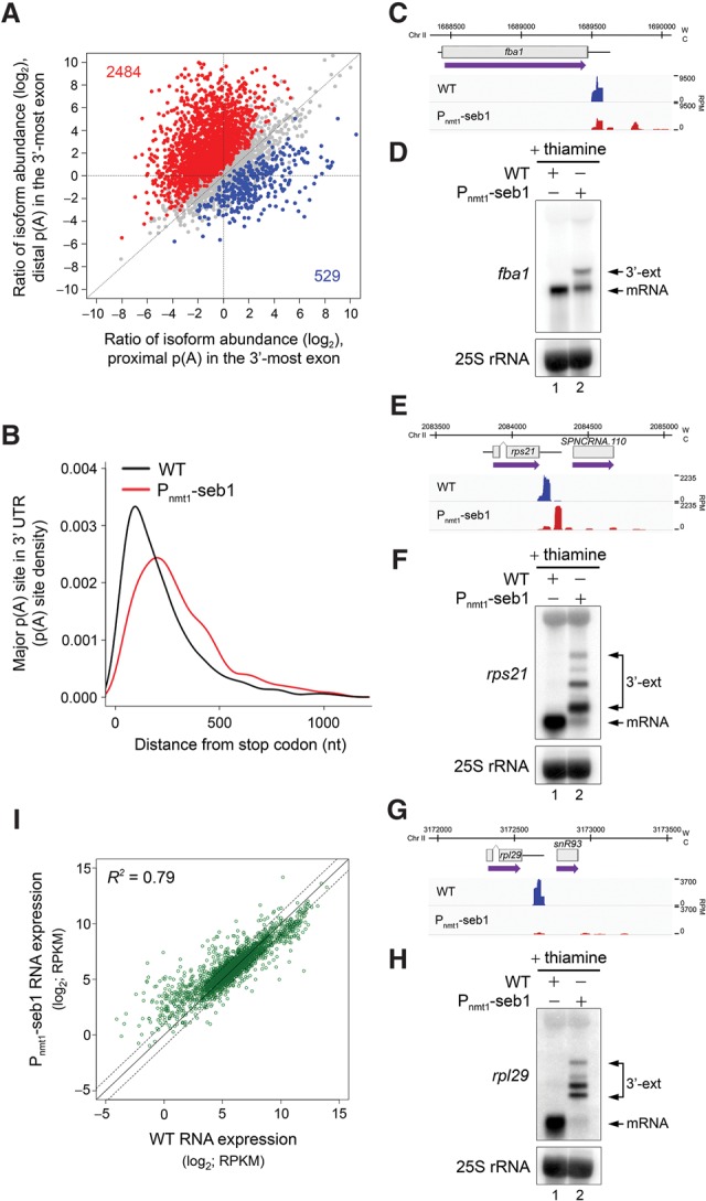
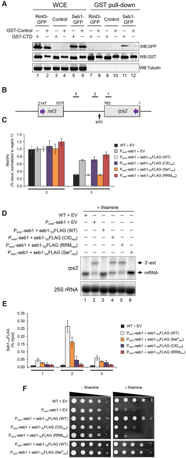
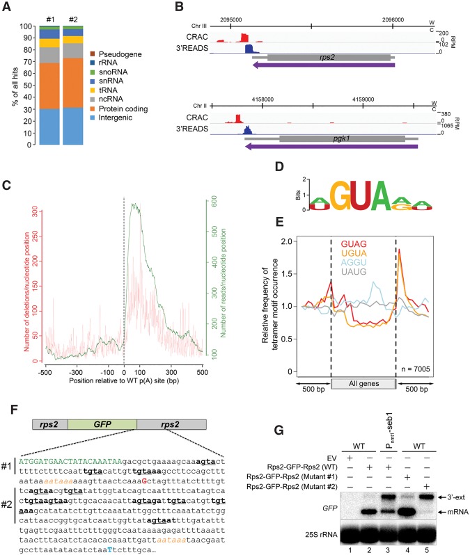
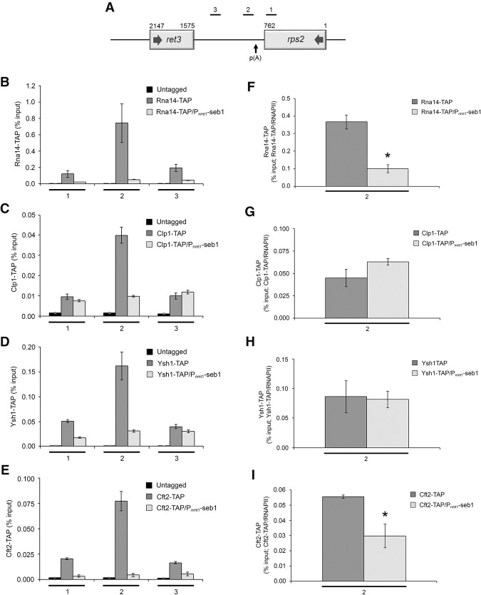
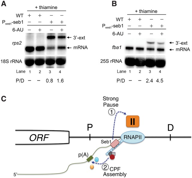
Termination of RNA polymerase II (RNAPII) transcription is associated with RNA 3' end formation. For coding genes, termination is initiated by the cleavage/polyadenylation machinery. In contrast, a majority of noncoding transcription events in Saccharomyces cerevisiae does not rely on RNA cleavage for termination but instead terminates via a pathway that requires the Nrd1-Nab3-Sen1 (NNS) complex. Here we show that the Schizosaccharomyces pombe ortholog of Nrd1, Seb1, does not function in NNS-like termination but promotes polyadenylation site selection of coding and noncoding genes. We found that Seb1 associates with 3' end processing factors, is enriched at the 3' end of genes, and binds RNA motifs downstream from cleavage sites. Importantly, a deficiency in Seb1 resulted in widespread changes in 3' untranslated region (UTR) length as a consequence of increased alternative polyadenylation. Given that Seb1 levels affected the recruitment of conserved 3' end processing factors, our findings indicate that the conserved RNA-binding protein Seb1 cotranscriptionally controls alternative polyadenylation.

Keywords: 3′ end processing; Nrd1; S. pombe; Seb1; polyadenylation; transcription termination.


Figures

Figure 1.
Figure 1.
Transcription termination defects in Seb1-depleted cells. (A) Schematic of the snR3 snoRNA locus. Bars above the gene show the positions of PCR products used for ChIP analyses in B and C. p(A) refers to the poly(A) site of the 3′ extended precursor (Lemay et al. 2010). (B) ChIP analyses of RNAPII density along the snR3 gene using extracts prepared from wild-type and the indicated mutant strains. ChIP signals (percent input) were normalized to region 1. Error bars indicate SD. n = 3 biological replicates from independent cell cultures. (C) ChIP analyses of HA-tagged Rpb3 (Rpb3-3XHA) along the snR3 gene in Seb1-depleted cells (Pnmt1-seb1) or control (wild-type) cells. Error bars indicate SD. n = 3 biological replicates from independent cell cultures. (D) Schematic showing the position of probes (1–5) used for TRO assays along the snR3 snoRNA locus. (E) Representative TRO blot for snR3. (FH) RNAPII profiles (ChIP-seq) across the snR3 (F), rps2 (G), and fba1 (H) genes for the indicated strains. (W) Watson strand; (C) Crick strand; (RPM) reads per million. (I) Cumulative RNAPII profile relative to poly(A) sites in the indicated strains. Curves show the sum of normalized ChIP-seq sequencing scores over a genomic region covering the major poly(A) site.
Figure 2.
Figure 2.
Seb1 interacts with the 3′ end processing machinery and is enriched at the 3′ ends of genes. (A) Coomassie blue staining of proteins copurified with Seb1-HTP (lane 2) and from a control untagged strain (lane 1). The arrow indicates the position of Seb1-HTP. (B) A subset of the top 10% of Seb1-associated proteins identified by liquid chromatography-tandem mass spectrometry (LC-MS/MS) is shown. The intensity represents the relative abundance (peptide intensity), while the percentage of coverage and the peptide number represent the unique peptide sequence coverage and the number of unique peptides, respectively. (C,D) Immunoblot analyses of whole-cell extracts (WCE) (lanes 1,2) and IgG-sepharose precipitates ([IP] IgG) (lanes 3–5) prepared from control Seb1-Myc cells or Seb1-Myc cells coexpressing a TAP tag version of Ysh1 (C) or Cft2 (D). (Lanes 4,5) Purification experiments were performed in the absence or presence of the benzonase nuclease. (EG) ChIP-seq analysis of Seb1-HTP occupancy along the snR3 (E), fba1 (F), and rps2 (G) genes. (W) Watson strand; (C) Crick strand; (RPM) reads per million. (H) Heat map of Seb1 DNA-binding sites derived from ChIP-seq for all RNAPII transcribed genes. Genes were sorted by length and aligned at their transcription start sites (TSSs). The curved line represents the poly(A) sites. Strength of binding is coded from white (no binding) to dark blue (strong binding).
Figure 3.
Figure 3.
Seb1 levels affect poly(A) site selection. (A) Regulation of APA site utilization in the 3′-most exon as determined by 3'READS. The number of genes with a significantly lengthened 3′ UTR (red dots) and the number of genes with a significantly shortened 3′ UTR (blue dots) are indicated in the graph. Significantly regulated isoforms are those with a P-value of <0.05 (Fisher's exact test). Only the two most abundant isoforms for each gene were analyzed. (B) Distribution of 3'READS-derived poly(A) sites relative to the upstream stop codon. The major poly(A) site is the one with the highest number of reads per gene. Mapped poly(A) sites in Seb1-depleted cells, as compared with wild-type cells, are, on average, significantly more distant from the stop codon. P-value < 2.22−16 by Wilcoxon rank-sum test. (CH) 3'READS profiles and Northern blot analyses of fba1 (C,D), rps21 (E,F), and rpl29 (G,H) genes in the indicated strains. (W) Watson strand; (C) Crick strand; (RPM) reads per million. 3′ extended transcripts that accumulate in the Seb1-depleted condition are shown (3′-ext). (I) RNA expression changes for transcripts with 3’ extended 3′ UTRs in wild-type and Seb1-depleted cells as measured by RNA-seq. (RPKM) Reads per kilobase per million. The coefficient of determination (R2) is indicated.
Figure 4.
Figure 4.
Seb1 requires functional CID and RRM domains for accurate 3′ end processing and transcription termination. (A) Immunoblot analysis of whole-cell extracts (WCE) (lanes 1–6) and glutathione-sepharose pull-downs (lanes 7–12) prepared from the indicated strains expressing either GST-CTD (odd-numbered lanes) or a control GST fusion protein (even-numbered lanes). (B) Bars above the rps2 gene show the positions of PCR products used for ChIP analyses. (C) RNAPII ChIP analysis using extracts prepared from the Pnmt1-seb1 conditional strain containing genomically integrated constructs that express the indicated versions of Flag-tagged Seb1 (wild type, CIDmut, Ser5mut, and RRMmut) (see the text; Supplemental Fig. S5a for description) as well as an empty control vector (EV). Cells were grown in the presence of thiamine to deplete endogenous Seb1. Error bars indicate SD. n = 3 biological replicates from independent cell cultures. (D) Northern blot analysis of rps2 mRNA from the indicated strains. The rps2 3′ extended transcripts are shown (3′-ext). (E) ChIP analyses of wild-type and mutant versions of Seb1-Flag along the rps2 gene. Control wild-type cells with an empty vector (black bars) were used as a negative control for the anti-Flag ChIP assays. Error bars indicate SD. n = 3 biological replicates from independent cell cultures. (F) Tenfold serial dilutions of the indicated strains were spotted on thiamine-free (left) or thiamine-containing (right) minimal medium.
Figure 5.
Figure 5.
Seb1 binds to GUA-containing motifs downstream from poly(A) sites. (A) Distribution of Seb1-bound reads between transcript classes for two independent CRAC experiments. (B) Seb1 CRAC cDNA read distribution (red) and 3'READS profile (blue) of rps2 and pgk1 genes in a wild-type strain. (W) Watson strand; (C) Crick strand; (RPM) reads per million. (C) Cumulative Seb1 RNA-binding sites relative to annotated poly(A) sites. The green curve (right Y-axis) shows the number of reads per nucleotide position, which is a measure of the binding preference. The red curve (left Y-axis) shows the number of deletions per nucleotide position, which is an indication of direct cross-linking. (D) Sequence logo of Seb1 cross-linking sites derived from the WebLogo application (Crooks et al. 2004) using the top 10 pyMotif-derived k-mers from each CRAC experiment. (E) Average gene distribution of tetrameric motifs derived from the Seb1 CRAC data (GUAG and UGUA) and control tetramers with shuffled dinucleotides (AGGU and UAUG). (F) Schematic of the rps2-GFP-rps2 construct used to address the functional significance of the Seb1 consensus motif in poly(A) site selection. Shown is a 405-nt region that includes the last seven codons of the GFP mRNA (in green) as well as the major poly(A) site of the GFP-rps2 mRNA detected in wild-type (G shown in red; +89 from stop codon) and Seb1-depleted (T shown in blue; +376 from stop codon) cells, as determined by 3′ RACE. The AAUAAA poly(A) signals are italicized in orange. Sequences in bold show Seb1 consensus motifs with the GUA core underlined. In mutant #1, the GUA core of the three Seb1 binding motifs located upstream of the wild-type rps2 cleavage site was mutated to CAC, whereas mutant #2 introduced CAC mutations in the eight Seb1-binding motifs located downstream from the rps2 cleavage site. (G) Northern blot analysis using total RNA prepared from wild-type (lanes 1,2,4,5) and Seb1-deficient (lane 3) cells that express either wild-type (lanes 2,3) or mutant (mutant #1 [lane 4] and mutant #2 [lane 5]) versions of the GFP-rps2 construct. Cells were grown in the presence of thiamine. The blot was analyzed using probes specific for the GFP mRNA and 25S rRNA.
Figure 6.
Figure 6.
Seb1 levels affect the cotranscriptional assembly of the cleavage/poly(A) machinery. (A) Bars above the rps2 gene show the positions of PCR products used for ChIP analyses. (BE) ChIP assays of TAP-tagged versions of Rna14 (B), Clp1 (C), Ysh1 (D), and Cft2 (E) in wild-type and Seb1-depleted cells (Pnmt1-seb1). An untagged control strain was used to monitor the background signal of the ChIP assays. (FI) Recruitment of 3′ end processing factors as a ratio of total RNAPII at the 3′ end of rps2 (region 2). ChIP signal of TAP-tagged 3′ end processing factors was divided by the total RNAPII signal at region 2. Region 2 was analyzed because it represents the location of maximal 3′ end processing factor recruitment. Error bars indicate SD. n = 3 biological replicates from independent cell cultures. (*) P < 0.05, Student's t-test.
Figure 7.
Figure 7.
Transcription kinetics contributes to Seb1-dependent poly(A) site selection. (A,B) Northern blot analysis of total RNA prepared from wild-type (lanes 1,2) and Seb1-depleted (lanes 3,4) cells that were treated (lanes 2,4) or not treated (lanes 1,3) with 6-AU. Blots were probed for rps2 (A) and fba1 (B) mRNAs. Ratios of proximal (P) relative to distal (D) mRNA isoforms are indicated (average from two independent experiments). (C) Model for Seb1-dependent poly(A) site selection. The passage of RNAPII through a poly(A) site is thought to induce a change in the kinetics of transcription elongation, including pausing of the RNAPII complex (Nag et al. 2006; Grosso et al. 2012; Davidson et al. 2014; Fusby et al. 2015; Nojima et al. 2015). We propose that the cooperative binding of Seb1 to the RNAPII CTD and to RNA motifs clustered downstream from poly(A) sites positively contributes to RNAPII pausing (1), thereby promoting poly(A) site recognition and assembly of a cleavage-competent cleavage/poly(A) (CPF) complex (2). In the absence of Seb1, RNAPII pausing is leaky, increasing the frequency of RNAPII complexes that reach distal (D) poly(A) sites.

Similar articles

Cited by

References

    1. Ahn SH, Kim M, Buratowski S. 2004. Phosphorylation of serine 2 within the RNA polymerase II C-terminal domain couples transcription and 3′ end processing. Mol Cell 13: 67–76. - PubMed
    1. Andersen PR, Domanski M, Kristiansen MS, Storvall H, Ntini E, Verheggen C, Schein A, Bunkenborg J, Poser I, Hallais M, et al. 2013. The human cap-binding complex is functionally connected to the nuclear RNA exosome. Nat Struct Mol Biol 20: 1367–1376. - PMC - PubMed
    1. Aranda A, Proudfoot NJ. 1999. Definition of transcriptional pause elements in fission yeast. Mol Cell Biol 19: 1251–1261. - PMC - PubMed
    1. Arigo JT, Eyler DE, Carroll KL, Corden JL. 2006. Termination of cryptic unstable transcripts is directed by yeast RNA-binding proteins Nrd1 and Nab3. Mol Cell 23: 841–851. - PubMed
    1. Bacikova V, Pasulka J, Kubicek K, Stefl R. 2014. Structure and semi-sequence-specific RNA binding of Nrd1. Nucleic Acids Res 42: 8024–8038. - PMC - PubMed

MeSH terms

Substances