. 2016 Aug 22;60(9):5563-72.
doi: 10.1128/AAC.00648-16. Print 2016 Sep.

Role of ABC and Solute Carrier Transporters in the Placental Transport of Lamivudine

Affiliations

Role of ABC and Solute Carrier Transporters in the Placental Transport of Lamivudine

Martina Ceckova et al. Antimicrob Agents Chemother. .

Abstract

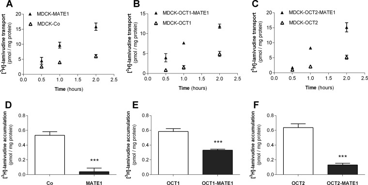
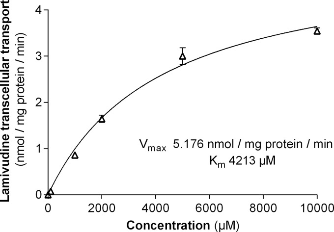
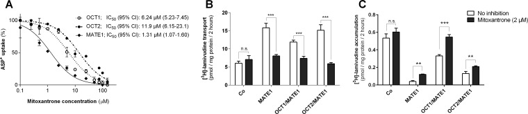
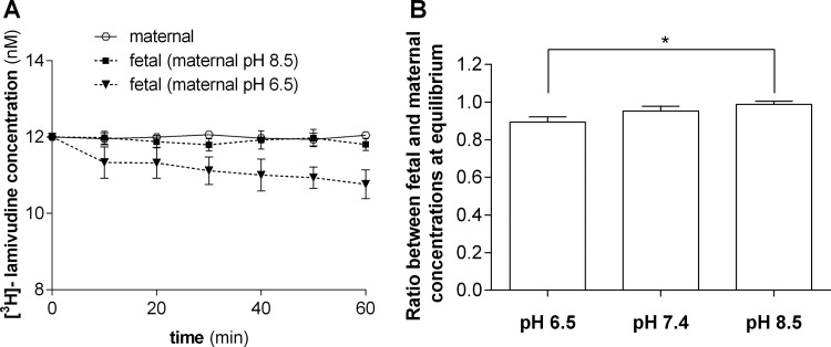
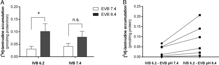
Lamivudine is one of the antiretroviral drugs of choice for the prevention of mother-to-child transmission (MTCT) in HIV-positive women. In this study, we investigated the relevance of drug efflux transporters P-glycoprotein (P-gp) (MDR1 [ABCB1]), BCRP (ABCG2), MRP2 (ABCC2), and MATE1 (SLC47A1) for the transmembrane transport and transplacental transfer of lamivudine. We employed in vitro accumulation and transport experiments on MDCK cells overexpressing drug efflux transporters, in situ-perfused rat term placenta, and vesicular uptake in microvillous plasma membrane (MVM) vesicles isolated from human term placenta. MATE1 significantly accelerated lamivudine transport in MATE1-expressing MDCK cells, whereas no transporter-driven efflux of lamivudine was observed in MDCK-MDR1, MDCK-MRP2, and MDCK-BCRP monolayers. MATE1-mediated efflux of lamivudine appeared to be a low-affinity process (apparent Km of 4.21 mM and Vmax of 5.18 nmol/mg protein/min in MDCK-MATE1 cells). Consistent with in vitro transport studies, the transplacental clearance of lamivudine was not affected by P-gp, BCRP, or MRP2. However, lamivudine transfer across dually perfused rat placenta and the uptake of lamivudine into human placental MVM vesicles revealed pH dependency, indicating possible involvement of MATE1 in the fetal-to-maternal efflux of the drug. To conclude, placental transport of lamivudine does not seem to be affected by P-gp, MRP2, or BCRP, but a pH-dependent mechanism mediates transport of lamivudine in the fetal-to-maternal direction. We suggest that MATE1 might be, at least partly, responsible for this transport.


Figures

FIG 1
FIG 1
Transport of lamivudine in single-transfected MDCK cells overexpressing OCT1, OCT2, and MATE1, double-transfected MDCK-cells overexpressing OCT1 or OCT2 and MATE1 (MDCK-OCT1-MATE1, MDCK-OCT2-MATE1), and vector control cells (MDCK-Co). Cells were seeded on Transwell semipermeable supports dividing a basal compartment and an apical compartment. Lamivudine (100 nM) was added to the basal compartment and sampled at time points 0.5, 1, and 2 h from the apical side of monolayers (A, B, C). Intracellular accumulation of lamivudine in the monolayers was determined in cell lysates at the end of the experiment (D, E, F). Data were analyzed by Student′s t test (***, P < 0.001 versus respective control [Co], OCT1, or OCT2) and are shown as means ± SD (n ≥ 3).
FIG 2
FIG 2
Concentration-dependent net transcellular transport of lamivudine by MATE1. Basolateral-to-apical transport of lamivudine across MDCK-MATE1 and MDCK-Co cells cultured as monolayers on Transwell membranes was investigated for increasing concentrations of nonradiolabeled lamivudine (1 × 10⁻⁴, 1 × 10⁻³, 0.01, 0.1, 1.0, 2.0, 5.0, and 10 mM) with addition of tracer [3H]lamivudine (16.7 nM) applied to the basolateral compartment. The transcellular transport of lamivudine across MDCK-Co monolayers was subtracted from that in MDCK-MATE1 cells at each concentration point. Kinetic parameters (Km and Vmax) were estimated by fitting MATE-specific transport rates to a Michaelis-Menten nonlinear equation. Data (nanomoles per milligram of protein per minute) represent the mean ± SD from three independent experiments.
FIG 3
FIG 3
Inhibitory effect of mitoxantrone on lamivudine transport by MATE1. (A) IC50s reflecting inhibition of ASP+ uptake into OCT1-, OCT2-, and MATE1-expressing MDCK cells by mitoxantrone. The IC50s with 95% confidential intervals were calculated from three independent measurements. (B and C) Effect of 2 μM mitoxantrone on transcellular transport (B) and intracellular accumulation (C) of lamivudine (100 nM) in monolayers of MATE1-expressing and control cells. Data were analyzed by two-way ANOVA with multiple comparisons (**, P < 0.01, ***, P < 0.001, versus respective noninhibited controls) and are shown as means ± SD (n ≥ 3). n.s., not significant.
FIG 4
FIG 4
Effect of maternal pH on elimination of lamivudine from the fetal circulation. In the closed-circuit perfusion setup, both the fetal and maternal sides of the placenta were simultaneously infused with 12 nM [3H]lamivudine. The fetal pH was set to 7.4, whereas the pH in the maternal reservoir was set to 6.5, 7.4, or 8.5. The fetal perfusate was recirculated for 60 min, and then fetal and maternal concentrations of lamivudine were compared. (A) Lamivudine fetal concentration over a 60-min perfusion at pH 6.5 and 8.5 applied on the maternal site. (B) Final ratio between fetal and maternal concentrations at equilibrium showing statistically significant difference between ratios calculated for perfusions at pH 6.5 and pH 8.5 (*, P < 0.05, Kruskal-Wallis test), suggesting involvement of a proton-cation antiporter system in lamivudine transplacental transport. Data are presented as means ± SD (n ≥ 3).
FIG 5
FIG 5
Effect of H+ gradient on [3H]lamivudine uptake by MVM vesicles from human term placentas. MVM vesicles were prepared in intravesicular buffer (IVB) at pH 6.2 or 7.4. (A) One-minute uptake of [3H]lamivudine was examined in extravesicular buffer (EVB) containing 100 nM [3H]lamivudine at pH 7.4 or 8.4. (B) Paired measurements for pH 6.2 IVB MVM vesicles showing stimulation of [3H]lamivudine uptake in the presence of an increased pH gradient (change, 422% ± 276% [mean ± SD]; n = 6; P = 0.044, paired t test). Data are presented as means ± SD from experiments with 5 to 7 placentas. n.s., not significant.

Similar articles

Cited by

References

    1. UNAIDS. 2015. Fact sheet 2014. Global statistics 2014; UNAIDS, Geneva, Switzerland: http://www.unaids.org/en/media/unaids/contentassets/documents/factsheet/....
    1. WHO. 2013. Consolidated guidelines on the use of antiretroviral drugs for treating and preventing HIV infection: recommendations for a public health approach. World Health Organization, Geneva, Switzerland. - PubMed
    1. Panel on Treatment of HIV-Infected Pregnant Women and Prevention of Perinatal Transmission. 2015. Recommendations for use of antiretroviral drugs in pregnant HIV-1-infected women for maternal health and interventions to reduce perinatal HIV transmission in the United States. http://aidsinfo.nih.gov/contentfiles/lvguidelines/PerinatalGL.pdf Accessed 26 May 2016.
    1. Lamberth JR, Reddy SC, Pan JJ, Dasher KJ. 2015. Chronic hepatitis B infection in pregnancy. World J Hepatol 7:1233–1237. doi:10.4254/wjh.v7.i9.1233. - DOI - PMC - PubMed
    1. Wong F, Pai R, Van Schalkwyk J, Yoshida EM. 2014. Hepatitis B in pregnancy: a concise review of neonatal vertical transmission and antiviral prophylaxis. Ann Hepatol 13:187–195. - PubMed

Substances

LinkOut - more resources