. 2016 Jul 1;61(1):33-41.
doi: 10.2144/000114435. eCollection 2016.

A phased strategy to differentiate human CD14+monocytes into classically and alternatively activated macrophages and dendritic cells

Affiliations

A phased strategy to differentiate human CD14+monocytes into classically and alternatively activated macrophages and dendritic cells

Jelani C Zarif et al. Biotechniques. .

Abstract

There are currently several in vitro strategies to differentiate human CD14(+) monocytes isolated from peripheral blood mononuclear cells (PBMCs) into the M1 or M2 macrophage cell types. Each cell type is then verified using flow cytometric analysis of cell-surface markers. Human CD14(+) monocytes have the potential to differentiate into M1 and M2 macrophages, both of which demonstrate varying degrees of cell-surface antigen overlap. Using multiple surface markers with current macrophage polarization protocols, our data reveal several limitations of currently used methods, such as highly ambiguous cell types that possess cell-surface marker overlap and functional similarities. Utilizing interleukin-6 (IL-6) and two phases of cytokine exposure, we have developed a protocol to differentiate human monocytes into M1, M2, or dendritic cells (DCs) with greater efficiency and fidelity relative to macrophages and DCs that are produced by commonly used methods. This is achieved via alterations in cytokine composition, dosing, and incubation times, as well as improvements in verification methodology. Our method reliably reproduces human in vitro monocyte-derived DCs and macrophage models that will aid in better defining and understanding innate and adaptive immunity, as well as pathologic states.

Keywords: M1 macrophages; M2 macrophages; cytokine; macrophage polarization; monocyte differentiation; monocyte-derived dendritic cells; multi-nucleated giant cells; peripheral blood mononuclear cells; phased strategy.


Conflict of interest statement

Competing interests statement: The authors declare no competing interest.

Figures

Figure 1
Figure 1. Schematic of Treatment strategies tested to drive monocyte differentiation into homogenous and functional myeloid cell types
Several polarization strategies were used (GM-CSF only, M-CSF only, continuous or phased). For polarization to the M1 phenotype, CD14+ PBMCs were given GM-CSF (20 ng/ml) for six days and then on day six cells were spiked with GM-CSF in concert with LPS, IL-6, and IFN-γ (20 ng/mL) for an additional four days. For M2 polarization, cells were treated with M-CSF (20 ng/mL) for six days and then on the sixth day, cells were then spiked with M-CSF in concert with IL-4, IL-6 and IL-13 (20 ng/mL) for an additional four days. Isolating human CD14+ monocytes and differentiating them into multiple myeloid cell types for 9 days, we demonstrate that timing, cytokine composition and dosage yields homogenous populations of macrophages and dendritic cells. CD14+ PBMCs under the phased cytokine treatment strategy were driven further than other strategies within the phenotypic continuum to produce homogenous populations that expressed canonical markers of their respective cell types. Under the phased strategy, M2 macrophages also displayed high mannose uptake and cellular fusion. Specific cytokine treatment on day 9 also increased moDC maturation as well as an increase in cell surface markers CD80, CD86, and CD86. We anticipate that the usage of the phased polarization method will be helpful for many immunology research groups as well as cancer researchers seeking to model the tumor microenvironment by incorporation human macrophages.
Figure 2
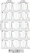
Figure 2. Changes in cell surface marker expression, cell size, and complexity of DCs, M1 and M2 macrophages after phased or constant cytokine treatments
Flow cytometric analysis of M1 and M2 macrophage cell surface markers expression for CD206 (A), CD36 (B), CD163 (C), E-Cadherin (Ecad) (D) and CD83 (E) under continuous or phased treatment regimens.
Figure 3
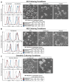
Figure 3. Successful polarization leads to increased surface expression of canonical M1, M2, and moDC cell surface markers
Panels (A–C) are separated into three sections. Histograms depict flow cytometric analysis of labeled surface markers with dashed lines depicting a representative unstained control. Accompanying tables represent the median fluorescent intensities (MFI) associated with the histograms. Values are shown as MFI relative to the unstained controls for that sample. Phase contrast images were taken of the cells prior to flow cytometric analysis. M1 surface markers analysis examining M1 markers, CD86 and CD80 are shown on the left, next to M2 markers, CD36 and IL-4Rα, on the right (A). M2 surface marker expression was determined for M2 markers CD206, CD163, E-Cadherin, and IL-4Rα (B). Expression levels for dendritic markers CD86, CD80, CD83 and CD1a were determined for moDCs (C).
Figure 4
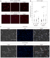
Figure 4. Polarized Macrophages are functional in vitro
M1 and M2 cells were treated with PBI-12-Man (10 μg/mL), a fluorescently tagged mannose for 30 min at 37°C to determine endocytosis. After time point was reached, both M1 and M2 cells were rinsed with 1X PBS, fixed, DAPI stained, mounted, and then visualized (A). Red fluorescence intensity per cell was quantified using the Metafer slide scanning software and presented as a box and whisker plot with *** representing p-values < 0.01. (B). M2 only and phased culturing conditions were grown for 14 days in culture, fixed, DAPI stained, mounted, and then visualized using phase contrast and fluorescence imaging (C). Individual multinucleated cells are outlined in the merged image with a red dashed line.

Similar articles

Cited by

References

    1. Ziegler-Heitbrock L, Ancuta P, Crowe S, Dalod M, Grau V, Hart DN, Leenen PJ, Liu YJ, et al. Nomenclature of monocytes and dendritic cells in blood. Blood. 2010;116(16):e74–80. - PubMed
    1. Geissmann F, Jung S, Littman DR. Blood monocytes consist of two principal subsets with distinct migratory properties. Immunity. 2003;19(1):71–82. - PubMed
    1. Jha AK, Huang SC, Sergushichev A, Lamproupoulou V, Ivanova Y, Loginicheva E, Chmielewski K, Stewart KM, et al. Network integration of parallel metabolic and transcriptional data reveals metabolic modules that regulate macrophage polarization. Immunity. 2015;42(3):419–30. - PubMed
    1. Biswas SK, Mantovani A. Macrophage plasticity and interaction with lymphocyte subsets: cancer as a paradigm. Nat Immunol. 2010;11(10):889–96. - PubMed
    1. Mantovani A, Sozzani S, Locati M, Allavena P, Siva A. Macrophage polarization: tumor-associated macrophages as a paradigm for polarized M2 mononuclear phagocytes. Trends Immunol. 2002;23(11):549–55. - PubMed

Publication types

LinkOut - more resources