. 2016 Aug;22(15-16):1006-15.
doi: 10.1089/ten.TEA.2016.0153. Epub 2016 Aug 1.

Three-Dimensional Mechanical Loading Modulates the Osteogenic Response of Mesenchymal Stem Cells to Tumor-Derived Soluble Signals

Affiliations

Three-Dimensional Mechanical Loading Modulates the Osteogenic Response of Mesenchymal Stem Cells to Tumor-Derived Soluble Signals

Maureen E Lynch et al. Tissue Eng Part A. 2016 Aug.

Abstract

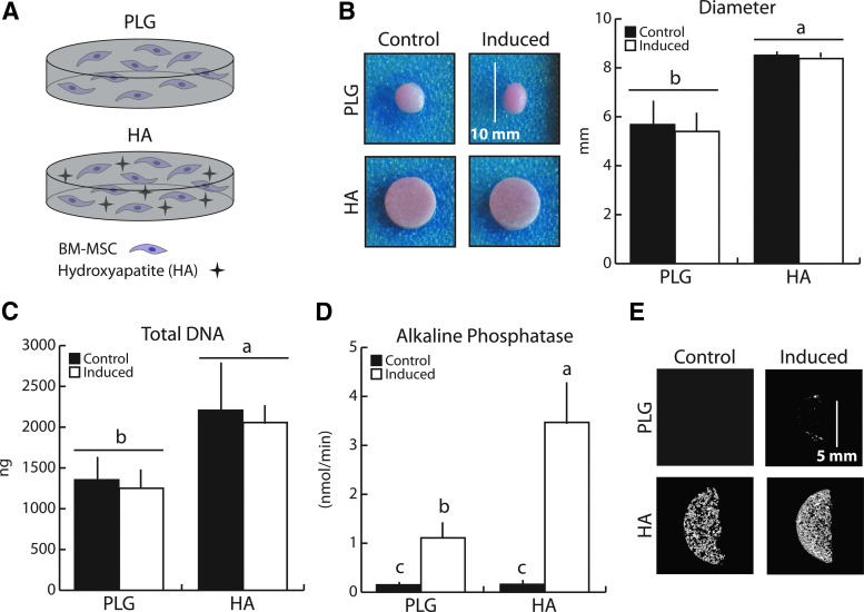
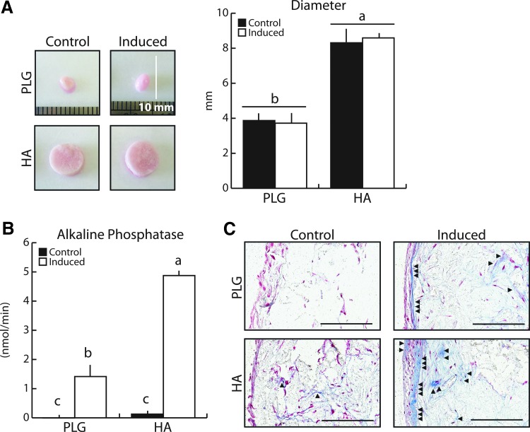
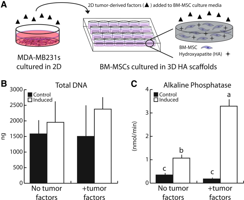
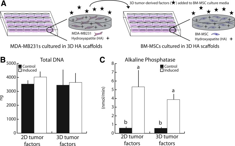
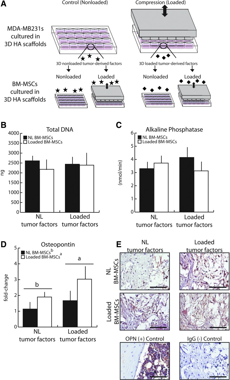
Dynamic mechanical loading is a strong anabolic signal in the skeleton, increasing osteogenic differentiation of bone marrow-derived mesenchymal stem cells (BM-MSCs) and increasing the bone-forming activity of osteoblasts, but its role in bone metastatic cancer is relatively unknown. In this study, we integrated a hydroxyapatite-containing three-dimensional (3D) scaffold platform with controlled mechanical stimulation to investigate the effects of cyclic compression on the interplay between breast cancer cells and BM-MSCs as it pertains to bone metastasis. BM-MSCs cultured within mineral-containing 3D poly(lactide-co-glycolide) (PLG) scaffolds differentiated into mature osteoblasts, and exposure to tumor-derived soluble factors promoted this process. When BM-MSCs undergoing osteogenic differentiation were exposed to conditioned media collected from mechanically loaded breast cancer cells, their gene expression of osteopontin was increased. This was further enhanced when mechanical compression was simultaneously applied to BM-MSCs, leading to more uniformly deposited osteopontin within scaffold pores. These results suggest that mechanical loading of 3D scaffold-based culture models may be utilized to evaluate the role of physiologically relevant physical cues on bone metastatic breast cancer. Furthermore, our data imply that cyclic mechanical stimuli within the bone microenvironment modulate interactions between tumor cells and BM-MSCs that are relevant to bone metastasis.


Figures

<b>FIG. 1.</b>
FIG. 1.
Mineral-containing scaffolds enhanced BM-MSC osteogenic differentiation after 21 days relative to control scaffolds. (A) To determine how the presence of HA affects osteogenic differentiation, mineral-free (PLG) and mineral-containing (HA) scaffolds were utilized to differentiate BM-MSCs (1.5 × 106 per scaffold) into osteoblasts. (B) HA scaffolds maintained their initial geometry after 21 days of culture, while control (PLG) scaffolds significantly contracted. (C) BM-MSCs growth in HA scaffolds was greater than that in PLG scaffolds and was independent of differentiation as detected by fluorimetric DNA analysis. (D) ALP enzyme activity increased with osteogenic induction and was enhanced in HA scaffolds. (E) Osteogenic induction resulted in increased mineral content as assessed by microCT, which was greater in HA scaffolds. Same letters (a, b, or c) indicate similar mean values, and groupings with different letters indicate that the difference is significant by post hoc comparison of means with Bonferroni correction (p < 0.05). ALP, alkaline phosphatase; BM-MSC, bone marrow-derived mesenchymal stem cell; HA, hydroxyapatite; BM-MSC, bone marrow-mesenchymal stem cell; PLG, poly(lactide-co-glycolide). Color images available online at www.liebertpub.com/tea
<b>FIG. 2.</b>
FIG. 2.
Long-term BM-MSC osteogenic differentiation in mineral-containing scaffolds verified the creation of a bone mimetic microenvironment. (A) After 60 days, HA-containing scaffolds continued to maintain their initial geometry, while PLG scaffolds continued to contract as confirmed by image analysis. (B) Heightened ALP enzyme activity with osteogenic induction was maintained in HA scaffolds. (C) Osteogenic differentiation resulted in greater Type I collagen deposition, as seen with Masson's Trichrome staining (collagen stains blue). The majority of matrix was deposited on scaffold surfaces; however, HA scaffolds also exhibited deposition within interior pores. Arrows indicate collagen deposition. Scale bars = 200 μm. Same letters (a, b, or c) indicate similar mean values, and groupings with different letters indicate that the difference is significant by post hoc comparison of means with Bonferroni correction (p < 0.05). Color images available online at www.liebertpub.com/tea
<b>FIG. 3.</b>
FIG. 3.
Human breast cancer-derived soluble factors increased BM-MSC osteogenic differentiation. (A) 2D tumor-conditioned media was collected from MDA-MB231 cultured on tissue culture plastic for 24 h in low serum media, after which media was collected, concentrated, and reconstituted with the appropriate BM-MSC culture media. (B) After 21 days, 2D tumor-conditioned media (+tumor factors) did not alter BM-MSC growth patterns as assessed by fluorimetric DNA analysis. (C) Tumor-derived factors enhanced ALP enzyme activity following induction, indicating increased osteogenic differentiation. Same letters (a, b, or c) indicate similar mean values, and groupings with different letters indicate that the difference is significant by post hoc comparison of means with Bonferroni correction (p < 0.05). 2D, two dimensional. Color images available online at www.liebertpub.com/tea
<b>FIG. 4.</b>
FIG. 4.
Human breast cancer-derived soluble factors generated in 2D versus 3D had similar effects on BM-MSC osteogenic differentiation. (A) 3D tumor-conditioned media was generated from MDA-MB231 tumor cells (1.5 × 106 per scaffold) cultured in HA-containing scaffolds for 24 h in low serum media, after which media was collected, concentrated, and reconstituted with the appropriate BM-MSC culture media. 2D media was generated as described previously. (B) After 21 days, BM-MSC growth patterns were similar when incubated in 2D versus 3D tumor-conditioned media (tumor factors) as indicated by fluorimetric DNA analysis. (C) Similarly, ALP enzyme activity was comparable between cultures supplemented with 2D and 3D TCM. Same letters (a, b, or c) indicate similar mean values, and groupings with different letters indicate that the difference is significant by post hoc comparison of means with Bonferroni correction (p < 0.05). 3D, three dimensional. Color images available online at www.liebertpub.com/tea
<b>FIG. 5.</b>
FIG. 5.
Mechanically loading tumor cells affected osteopontin gene expression and protein deposition in BM-MSCs. (A) Loaded and Nonloaded (NL) tumor-conditioned media were generated from MDA-MB231 tumor cells (1.5 × 106 per scaffold) cultured in HA-containing scaffolds that underwent 1 h of cyclic compression (1 Hz, 10% peak strain) and then processed as previously described. Control, nonloaded media was generated similarly. (B) Mechanical loading of tumor cells did not alter downstream growth patterns in BM-MSCs. (C) Similarly, no differences in ALP enzyme activity were detected. (D) Quantitative real-time polymerase chain reaction (qRT-PCR) revealed that mechanical loading increased OPN expression in BM-MSCs, and this effect was more pronounced when BM-MSCs were cultured in conditioned media from loaded tumor cells (Loaded tumor factors). (E) Immunohistochemistry suggested that mechanical loading also stimulated OPN protein deposition by BM-MSCs within scaffolds. Furthermore, BM-MSCs cultured with conditioned media from mechanically loaded versus nonloaded tumor cells resulted in more uniform protein deposition within the scaffolds. Scale bars = 100 μm. Same letters (a or b) indicate similar mean values, and groupings with different letters indicate that the difference is significant by post hoc comparison of means with Bonferroni correction (p < 0.05). Color images available online at www.liebertpub.com/tea

Similar articles

Cited by

References

    1. DeSantis C.E., Lin C.C., Mariotto A.B., Siegel R.L., Stein K.D., Kramer J.L., Alteri R., Robbins A.S., and Jemal A. Cancer treatment and survivorship statistics, 2014. CA Cancer J Clin 64, 252, 2014 - PubMed
    1. Harbeck N., Salem M., Nitz U., Gluz O., and Liedtke C. Personalized treatment of early-stage breast cancer: present concepts and future directions. Cancer Treat Rev 36, 584, 2010 - PubMed
    1. Coleman R.E. Bisphosphonates in breast cancer. Ann Oncol 16, 687, 2005 - PubMed
    1. Manolagas S.C. Birth and death of bone cells: basic regulatory mechanisms and implications for the pathogenesis and treatment of osteoporosis. Endocr Rev 21, 115, 2000 - PubMed
    1. Guise T.A. The vicious cycle of bone metastases. J Musculoskelet Neuronal Interact 2, 570, 2002 - PubMed