. 2017 Jun;37(6):2002-2012.
doi: 10.1177/0271678X16659302. Epub 2016 Jan 1.

Sustained diffusion reversal with in-bore reperfusion in monkey stroke models: Confirmed by prospective magnetic resonance imaging

Affiliations

Sustained diffusion reversal with in-bore reperfusion in monkey stroke models: Confirmed by prospective magnetic resonance imaging

Kyung Sik Yi et al. J Cereb Blood Flow Metab. 2017 Jun.

Abstract

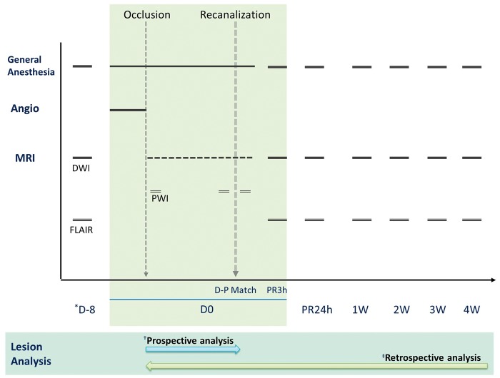
Although early diffusion lesion reversal after recanalization treatment of acute ischaemic stroke has been observed in clinical settings, the reversibility of lesions observed by diffusion-weighted imaging remains controversial. Here, we present consistent observations of sustained diffusion lesion reversal after transient middle cerebral artery occlusion in a monkey stroke model. Seven rhesus macaques were subjected to endovascular transient middle cerebral artery occlusion with in-bore reperfusion confirmed by repeated prospective diffusion-weighted imaging. Early diffusion lesion reversal was defined as lesion reversal at 3 h after reperfusion. Sustained diffusion lesion reversal was defined as the difference between the ADC-derived pre-reperfusion maximal ischemic lesion volume (ADCD-P Match) and the lesion on 4-week follow-up FLAIR magnetic resonance imaging. Diffusion lesions were spatiotemporally assessed using a 3-D voxel-based quantitative technique. The ADCD-P Match was 9.7 ± 6.0% (mean ± SD) and the final infarct was 1.2-6.0% of the volume of the ipsilateral hemisphere. Early diffusion lesion reversal and sustained diffusion lesion reversal were observed in all seven animals, and the calculated percentages compared with their ADCD-P Match ranged from 8.3 to 51.9% (mean ± SD, 26.9 ± 15.3%) and 41.7-77.8% (mean ± SD, 65.4 ± 12.2%), respectively. Substantial sustained diffusion lesion reversal and early reversal were observed in all animals in this monkey model of transient focal cerebral ischaemia.

Keywords: Animal model; brain imaging; brain ischaemia; brain recovery; diffusion-weighted magnetic resonance imaging.


Figures

Figure 1.
Figure 1.
Timeline of monkey stroke model using prospective repeated MRI and in-bore reperfusion. Baseline MRI was performed in all experimental animals with anaesthesia at 8 days before MCAO procedure to evaluate any underlying brain disease and to serve as a standard for comparison with the post-infarct status. After endovascular occlusion of the middle cerebral artery (MCAO), the experimental animals were transported to the MRI suite while maintaining endovascular MCAO. Post-occlusion diffusion MRIs (DWIs) were repeated every 10 ∼ 15 min thereafter (animals 1 ∼ 2, every 10 min; animals 3 ∼ 7, every 15 min). Transient right MCAO was maintained until the ADC-derived volume of the focal ischaemia had plateaued with variable duration to each animal (plateau was defined when the ischaemic lesions did not change for three consecutive DWI MRIs). Immediately prior to reperfusion, DSC perfusion MRI was performed to confirm diffusion–perfusion matching. Follow-up MRIs were performed at 3-h post-reperfusion (PR3h), 24 h and then every week for 4 weeks. (*D-8, 8 days before MCAO; Prospective analysis was performed using ImageJ software for real-time determination of an acute phase diffusion lesion after MCAO to determine the time point for arterial recanalization; Retrospective analysis was performed by a sophisticated processing method for precise lesional evaluation).
Figure 2.
Figure 2.
Steps of MR data processing and lesion segmentation (representative demonstration in animal 4). The following three processing steps were implemented for MR imaging processing. (1) Affine registration of the entire time series of ADC maps and FLAIRs to baseline FLAIR (normal brain parenchyma at 8 days before MCAO) using FLIRT in FSL; (2) lesion segmentation on ADC maps and FLAIRs using ITKsnap software (http://www.itksnap.org/pmwiki/pmwiki.php). (a) Conversion to relative ADC (rADC) map compared with the mean normal contralateral hemisphere. (b) Automatic thresholding (first step): lesion included voxels with value of rADC less than 80 (the colour-coded image shows a step of speed image generation in ITKsnap). (c) Semi-automatic thresholding (second step): fine adjustment (rADC = 78 in animal 4) compared with pre-reperfusion ADC map.
Figure 3.
Figure 3.
Coregistered serial MR images of experimental animals subjected to endovascular MCAO and in-bore reperfusion. Early DLR (eDLR) was defined as the ADC-derived hemispheric lesion volume (HLV) difference between the pre-reperfusion lesion (ADCD-P Match) and the lesion volume at 3 h after reperfusion (ADCPR3h), and sustained DLR (sDLR) was the volume difference between the ADCD-P Match and the lesion on the 4-week follow-up FLAIR MRI (FLAIR4w) after the procedure. Affine registration was applied to all images for intrasubject coregistration.
Figure 4.
Figure 4.
Temporal changes in mean abnormal lesion volumes of all experimental animals during 4 weeks of follow-up. Error bars present standard deviation of the mean; %HLV, percentage of the hemispheric lesion volume.
Figure 5.
Figure 5.
Diffusion lesion reversals in experimental animals. Ratio of eDLR and ratio of sDLR are presented (ratio of eDLR=eDLR/ADCD-P Match; ratio of sDLR=sDLR/ADCD-P Match).
Figure 6.
Figure 6.
Diffusion and perfusion MR images at the pre-reperfusion time point (animals 1, 4, 5, and 6). Representative MR images of ADC maps and DSC perfusion MRIs (CBV/CBF maps) showing diffusion–perfusion match for all animals.

References

    1. Berkhemer OA, Fransen PS, Beumer D, et al. A randomized trial of intraarterial treatment for acute ischemic stroke. N Engl J Med 2015; 372: 11–20. - PubMed
    1. Goyal M, Demchuk AM, Menon BK, et al. Randomized assessment of rapid endovascular treatment of ischemic stroke. N Engl J Med 2015; 372: 1019–1030. - PubMed
    1. Campbell BC, Mitchell PJ, Kleinig TJ, et al. Endovascular therapy for ischemic stroke with perfusion-imaging selection. N Engl J Med 2015; 372: 1009–1018. - PubMed
    1. Guadagno JV, Warburton EA, Jones PS, et al. The diffusion-weighted lesion in acute stroke: heterogeneous patterns of flow/metabolism uncoupling as assessed by quantitative positron emission tomography. Cerebrovasc Dis 2005; 19: 239–246. - PubMed
    1. Gonzalez RG, Copen WA, Schaefer PW, et al. The Massachusetts General Hospital acute stroke imaging algorithm: an experience and evidence based approach. J Neurointerv Surg 2013; 5 Suppl 1: i7–i12. - PMC - PubMed

LinkOut - more resources