. 2016 Jul 12:7:12140.
doi: 10.1038/ncomms12140.

Dynamic polarization vision in mantis shrimps

Affiliations

Dynamic polarization vision in mantis shrimps

Ilse M Daly et al. Nat Commun. .

Abstract

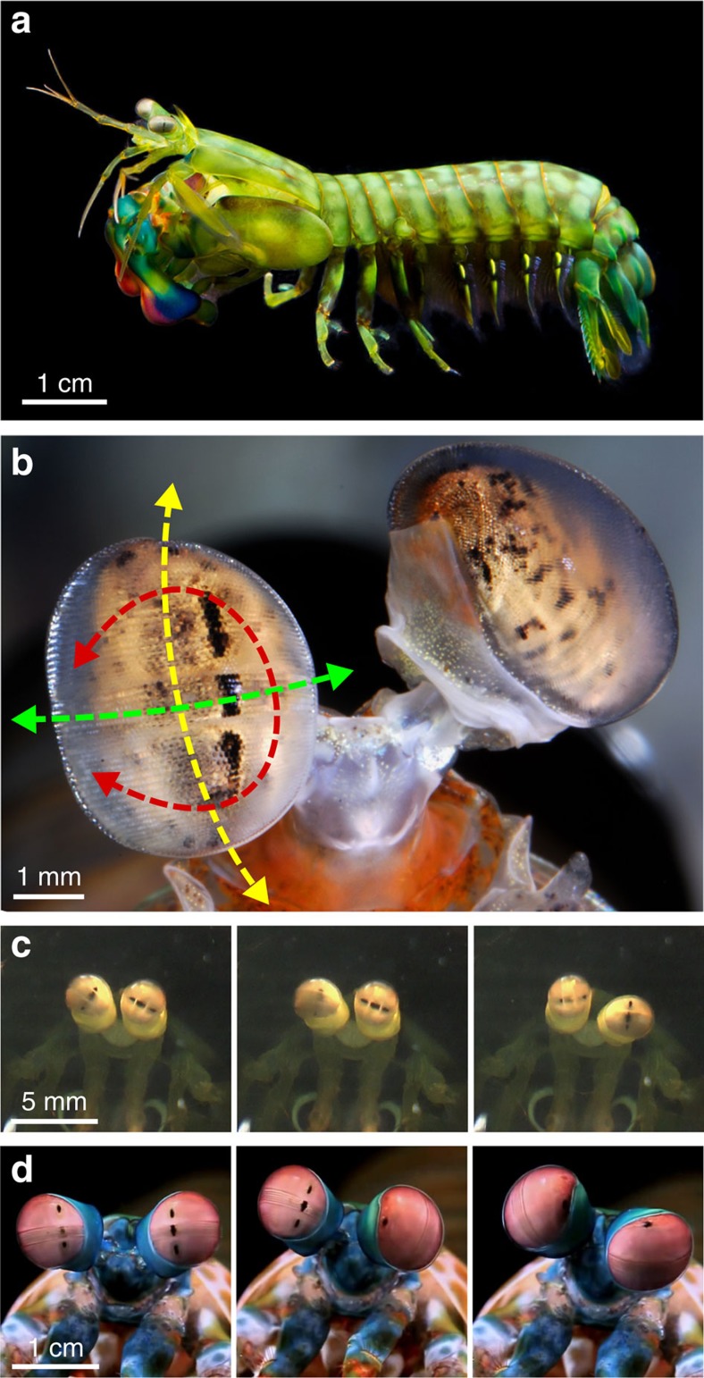
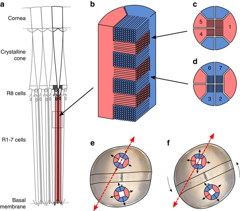
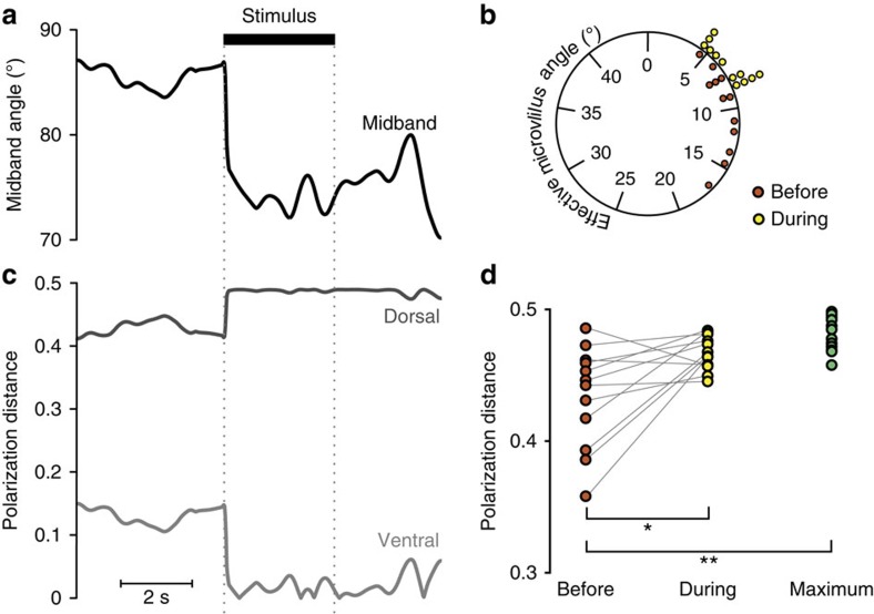
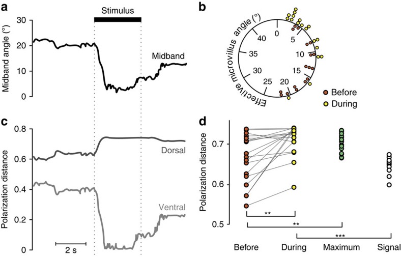
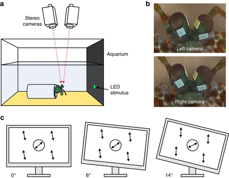
Gaze stabilization is an almost ubiquitous animal behaviour, one that is required to see the world clearly and without blur. Stomatopods, however, only fix their eyes on scenes or objects of interest occasionally. Almost uniquely among animals they explore their visual environment with a series pitch, yaw and torsional (roll) rotations of their eyes, where each eye may also move largely independently of the other. In this work, we demonstrate that the torsional rotations are used to actively enhance their ability to see the polarization of light. Both Gonodactylus smithii and Odontodactylus scyllarus rotate their eyes to align particular photoreceptors relative to the angle of polarization of a linearly polarized visual stimulus, thereby maximizing the polarization contrast between an object of interest and its background. This is the first documented example of any animal displaying dynamic polarization vision, in which the polarization information is actively maximized through rotational eye movements.


Figures

Figure 1
Figure 1. Mantis shrimp eye movements.
(a) Side view of a Gonodactylus smithii. (b) Rotational degrees of freedom of stomatopod eyes relative to the external environment, as demonstrated in Odontodactylus cultrifer. Yellow arrows=pitch (up–down); green arrows=yaw (side-to-side); red arrows=torsional (roll) rotations. The midband is visible as a distinct stripe of ommatidial facets dividing the eye into dorsal and ventral hemispheres. (c,d) Series of video still frames demonstrating the torsional rotation range in G. smithii (c) and Odontodactylus scyllarus (d). (c) left eye - 45°, 85°, 0°; right eye - 30°, 20°, 90°; and (d) left eye - 90°, 80°, 0°; right eye - 90°, 0°, 90°.
Figure 2
Figure 2. Diagrams illustrating the polarization anatomy of a mantis shrimp eye and the relevant geometries.
(a) Longitudinal section through a stomatopod eye hemisphere showing the main rhabdom (retinular (R) cells 1–7) and the distal R8 cell (redrawn from Marshall et al.26). (b) Bi-directional microvillar projections from two retinular cells (coloured red and blue for illustrative purposes) forming stacked layers within the main rhabdom (similar to standard crustacean eye anatomy). (c,d) the orientation of microvilli in alternate layers of the main rhabdom. (e) Relative orientations of microvilli in the main rhabdoms in each hemisphere (note that these are not drawn to scale). There is a 45° skew in the overall orientation of the microvillar directions between the dorsal and ventral hemispheres. In e, the dorsal microvilli are optimally aligned for detecting the incoming polarized light stimulus (dashed red arrow; that is, effective angle ϕ=0°). In f, the eye has rotated by 22.5° with the consequence that both sets of orthogonal microvilli are maximally misaligned with the stimulus (that is, ϕ=22.5°). (bd) Redrawn from Goldsmith. Background image in e and f adapted from a photo by R Caldwell.
Figure 3
Figure 3. Experimental results for the LED experiment.
(a) An example of the torsional rotation of the midband in response to the polarized LED stimulus (onset and offset of stimulus is denoted by black bar and dotted grey lines). (b) Angle subtended between the stimulus angle of polarization and the nearest polarization receptor before the onset (red circles) and during (yellow circles) the stimulus presentation. (c) An example of the polarization distance (PD) calculated for the dorsal (grey line) and ventral (light grey line) pairs of polarization receptors during a single stimulus presentation. (d) Same data as b, with paired comparison between the calculated PD, both before (red circles), and during (yellow circles) the stimulus presentation and compared with a set of bootstrap resampled maximum data points (green circles). Stars represent levels of statistical significance: *P<0.05; **P<0.01; ***P<0.001.
Figure 4
Figure 4. Experimental results where six O. scyllarus were presented with expanding circular looming stimuli displayed on an LCD monitor.
Captions as for the LED experiment. Note that the angle measured in b is between the optimal receptor alignment for detecting the maximum polarization contrast, and the nearest set of polarization receptors. (d) Same data as b, with paired comparison between the calculated PD, both before (red circles), and during (yellow circles) the stimulus presentation and compared with a set of bootstrap resampled maximum data points (green circles). In addition, the modelled data set representing the viewed PD if the closest group of microvilli were aligned with the stimulus angle of polarization (open circles) is shown. Stars represent levels of statistical significance: *P<0.05; **P<0.01; ***P<0.001.
Figure 5
Figure 5. Experimental method.
(a) Side view of the experimental apparatus for the LED experiment. (b) Frame-grabs from each video camera at a single point in time showing the pair of tracking markers fixed to each eye-stalk. (c) Illustration of the tilt positions of the modified LCD monitor for the second experimental setup and the consequent effect on loom and background angle of polarization.

Similar articles

Cited by

References

    1. Land M. F. Motion and vision: why animals move their eyes. J. Comp. Physiol. A 185, 341–352 (1999). - PubMed
    1. Land M. F. Head movement of flies during visually guided flight. Nature 243, 299–300 (1973).
    1. Troje N. F. & Frost B. J. Head-bobbing in pigeons: how stable is the hold phase? J. Exp. Biol. 203, 935–940 (2000). - PubMed
    1. Zeil J. & Hemmi J. M. The visual ecology of fiddler crabs. J. Comp. Physiol. A 192, 1–25 (2006). - PubMed
    1. Layne J. E., Wicklein M., Dodge F. A. & Barlow R. B. Prediction of maximum allowable retinal slip speed in the fiddler crab Uca pugilator. Biol. Bull 193, 202–203 (1997). - PubMed

Publication types

LinkOut - more resources