

Association of epithelial-mesenchymal transition and nuclear cofilin with advanced urothelial cancer
- PMID: 27402302
- PMCID: PMC5629972
- DOI: 10.1016/j.humpath.2016.06.020
Association of epithelial-mesenchymal transition and nuclear cofilin with advanced urothelial cancer
Abstract
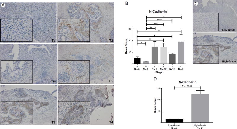
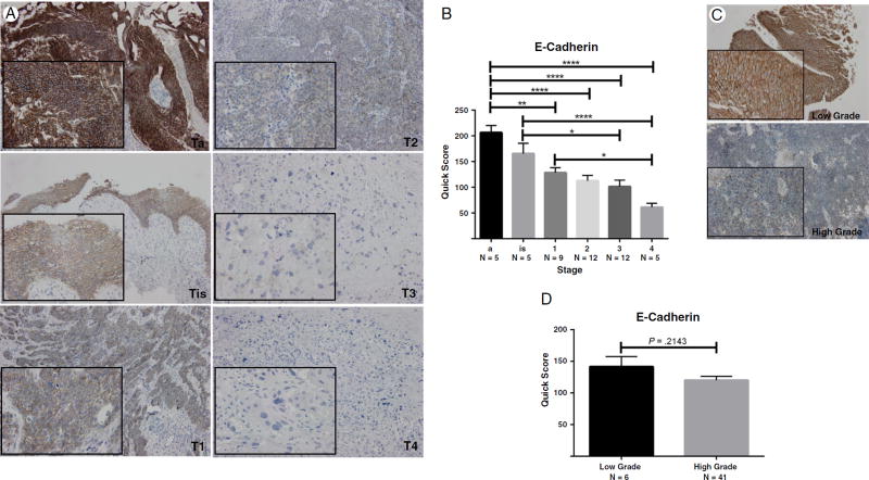
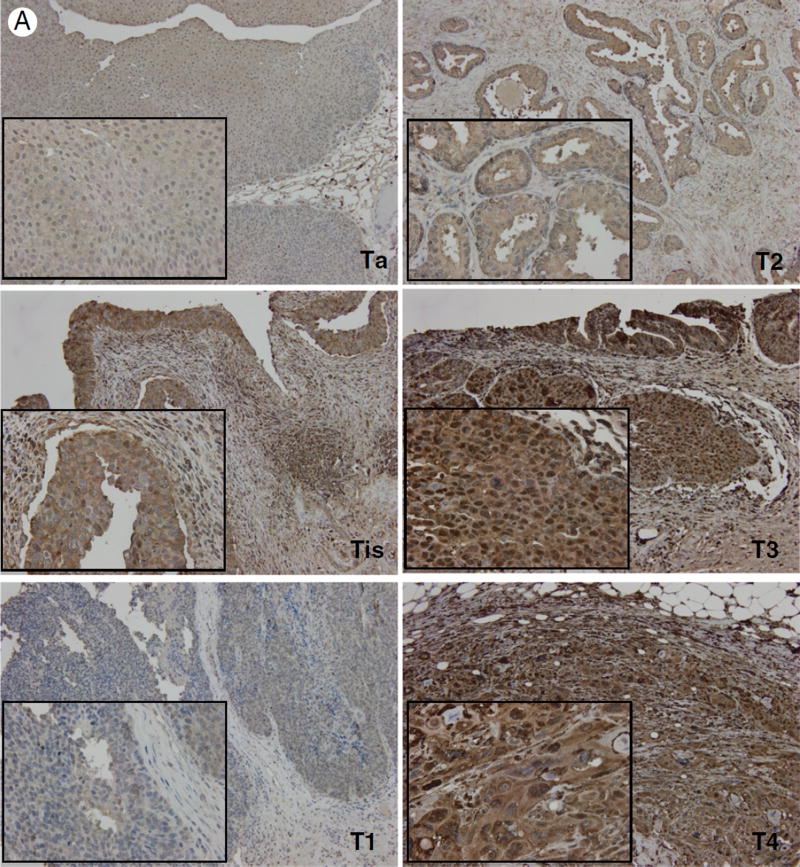
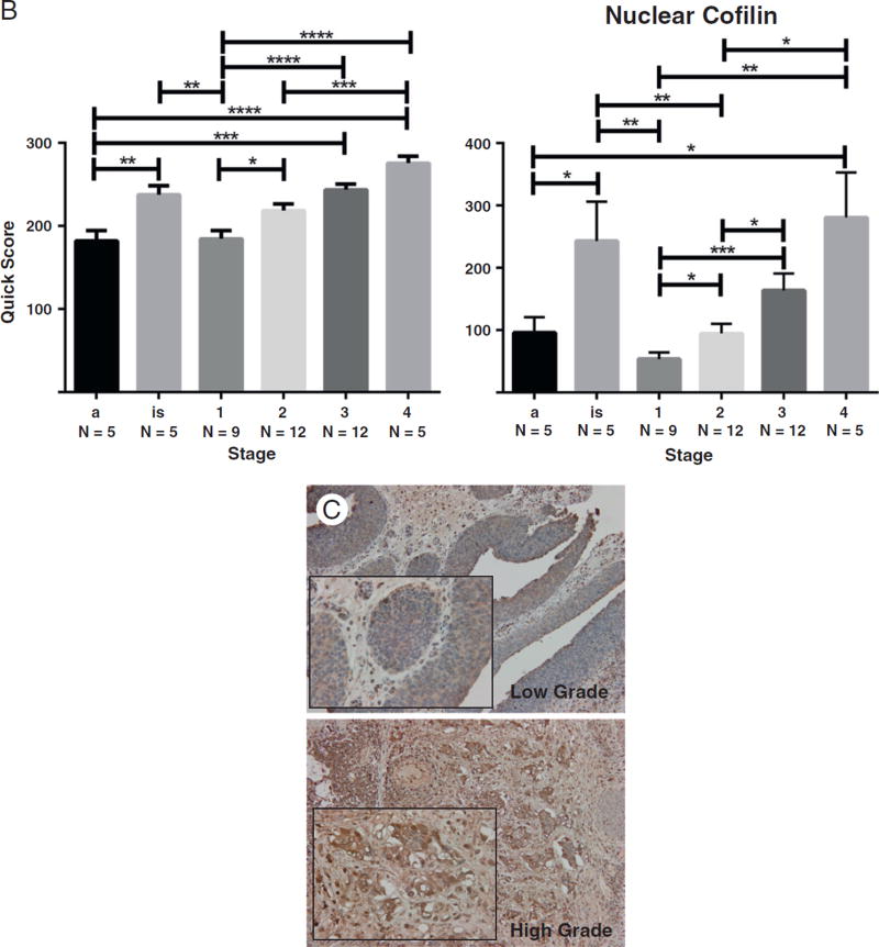
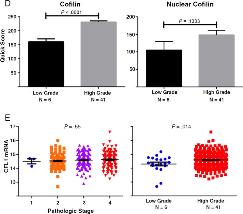
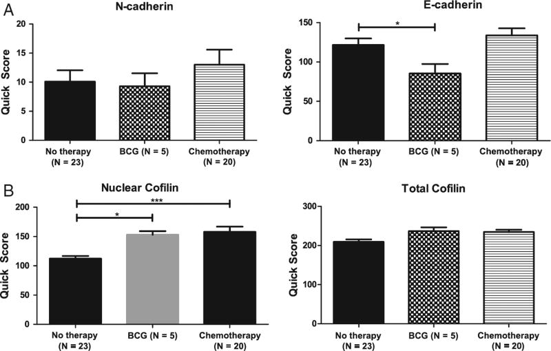
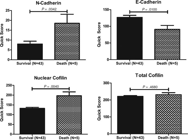
Tumor epithelial cells undergo a morphologic shift through the process of EMT with characteristic loss of cell polarity, conferring invasive and metastatic properties during cancer progression. Signaling by transforming growth factor-β mediates EMT programming and its phenotypic reversal to mesenchymal-epithelial transition. The role of EMT in bladder cancer progression to advanced disease is poorly understood. In this study, we conducted a retrospective analysis of the EMT landscape and actin cytoskeleton remodeling in a series of human bladder cancer specimens. Immunoreactivity for E-cadherin, N-cadherin, and vimentin protein expression was performed toward establishing an EMT signature in human bladder cancer. Serial sections were assessed for the primary regulator of the actin cytoskeleton remodeling and transforming growth factor-β signaling effector, cofilin. Our results demonstrate that EMT induction in clinical bladder cancer specimens is significantly associated with bladder cancer progression to high-grade, invasive disease. Evaluation of expression and cellular localization of the cytoskeleton regulator cofilin revealed a significant association between overexpression of nuclear cofilin with bladder cancer progression. This study is of translational significance in defining the value of EMT signature and cytoskeletal cofilin as potential tumor markers and targetable platforms for the treatment of invasive bladder cancer.
Keywords: Actin cytoskeleton; Bladder cancer; E-cadherin; N-cadherin; Phenotypic changes.
Copyright © 2016 Elsevier Inc. All rights reserved.
Conflict of interest statement
Competing Interest: The authors declare no potential conflict.
Figures
Similar articles
-
Cofilin-1 signaling mediates epithelial-mesenchymal transition by promoting actin cytoskeleton reorganization and cell-cell adhesion regulation in colorectal cancer cells.Biochim Biophys Acta Mol Cell Res. 2019 Mar;1866(3):418-429. doi: 10.1016/j.bbamcr.2018.10.003. Epub 2018 Oct 5. Biochim Biophys Acta Mol Cell Res. 2019. PMID: 30296500
-
WHO 1973 grade 3 and infiltrative growth pattern proved, aberrant E-cadherin expression tends to be of predictive value for progression in a series of stage T1 high-grade bladder cancer after organ-sparing approach.Int Urol Nephrol. 2017 Mar;49(3):431-437. doi: 10.1007/s11255-016-1491-9. Epub 2016 Dec 29. Int Urol Nephrol. 2017. PMID: 28035618
-
Expression profile of epithelial-mesenchymal transition markers in non-muscle-invasive urothelial carcinoma of the bladder: correlation with intravesical recurrence following transurethral resection.Urol Oncol. 2015 Mar;33(3):110.e11-8. doi: 10.1016/j.urolonc.2014.08.012. Epub 2014 Sep 26. Urol Oncol. 2015. PMID: 25262382
-
Urothelial cancer stem cells and epithelial plasticity: current concepts and therapeutic implications in bladder cancer.Cancer Metastasis Rev. 2015 Dec;34(4):691-701. doi: 10.1007/s10555-015-9589-6. Cancer Metastasis Rev. 2015. PMID: 26328525 Review.
-
Role of epithelial-to-mesenchymal transition (EMT) in drug sensitivity and metastasis in bladder cancer.Cancer Metastasis Rev. 2009 Dec;28(3-4):335-44. doi: 10.1007/s10555-009-9194-7. Cancer Metastasis Rev. 2009. PMID: 20012924 Free PMC article. Review.
Cited by
-
Cofilin-1 levels and intracellular localization are associated with melanoma prognosis in a cohort of patients.Oncotarget. 2018 May 8;9(35):24097-24108. doi: 10.18632/oncotarget.25303. eCollection 2018 May 8. Oncotarget. 2018. PMID: 29844875 Free PMC article.
-
Quantitative nuclear histomorphometric features are predictive of Oncotype DX risk categories in ductal carcinoma in situ: preliminary findings.Breast Cancer Res. 2019 Oct 17;21(1):114. doi: 10.1186/s13058-019-1200-6. Breast Cancer Res. 2019. PMID: 31623652 Free PMC article.
-
Predictive value of phenotypic signatures of bladder cancer response to cisplatin-based neoadjuvant chemotherapy.Urol Oncol. 2019 Sep;37(9):572.e1-572.e11. doi: 10.1016/j.urolonc.2019.06.020. Epub 2019 Jul 17. Urol Oncol. 2019. PMID: 31326313 Free PMC article.
-
miR-182-5p affects human bladder cancer cell proliferation, migration and invasion through regulating Cofilin 1.Cancer Cell Int. 2019 Feb 28;19:42. doi: 10.1186/s12935-019-0758-5. eCollection 2019. Cancer Cell Int. 2019. PMID: 30858759 Free PMC article.
-
The Role of Actin Dynamics and Actin-Binding Proteins Expression in Epithelial-to-Mesenchymal Transition and Its Association with Cancer Progression and Evaluation of Possible Therapeutic Targets.Biomed Res Int. 2018 Jan 16;2018:4578373. doi: 10.1155/2018/4578373. eCollection 2018. Biomed Res Int. 2018. PMID: 29581975 Free PMC article. Review.
References
-
- Siegel R, Miller K, Jemal A. Cancer statistics, 2015. Cancer J Clin. 2015;65:5–29. - PubMed
-
- Ro JY, Staerkel GA, Ayala AG. Cytologic and histologic features of superficial bladder cancer. Urol Clin North Am. 1992;19:435–453. - PubMed
-
- Holmang S, Hedelin H, Anderstrom C, Johansson SL. The relationship among multiple recurrences, progression and prognosis of patients with stages Ta and T1 transitional cell cancer of the bladder followed for at least 20 years. J Urol. 1995;153:1823–1826. - PubMed
-
- Morgan TM, Clark PE. Bladder cancer. Curr Opin Oncol. 2010;22:242–249. - PubMed
-
- Abida W, Bajorin DF, Rosenberg JE. First-line treatment and prognostic factors of metastatic bladder cancer for platinum-eligible patients. Hematol Oncol Clin North Am. 2015;29:319–328. - PubMed
Publication types
MeSH terms
Substances
Grants and funding
LinkOut - more resources
Full Text Sources
Other Literature Sources
Medical
Research Materials
Miscellaneous
