. 2016 Aug 1;311(2):H433-44.
doi: 10.1152/ajpheart.00176.2016. Epub 2016 Jul 8.

Stimulation of the hypothalamic arcuate nucleus increases brown adipose tissue nerve activity via hypothalamic paraventricular and dorsomedial nuclei

Affiliations

Stimulation of the hypothalamic arcuate nucleus increases brown adipose tissue nerve activity via hypothalamic paraventricular and dorsomedial nuclei

Vineet C Chitravanshi et al. Am J Physiol Heart Circ Physiol. .

Abstract

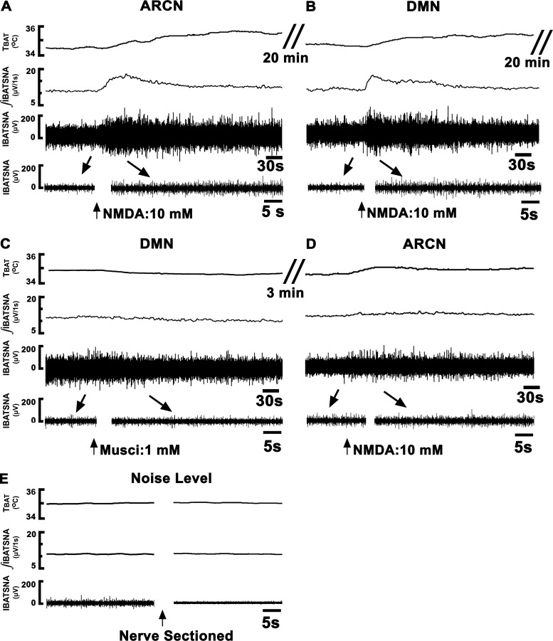
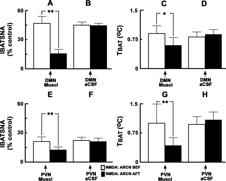
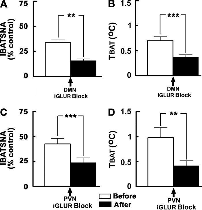
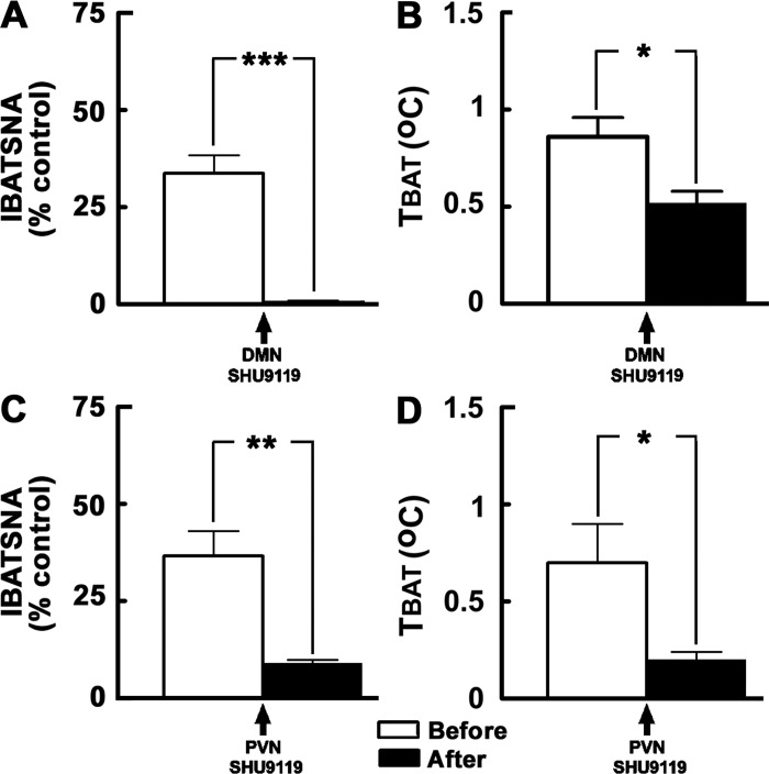
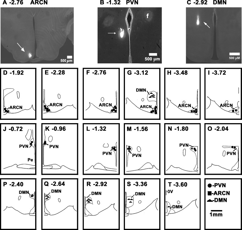
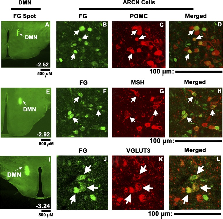
Hypothalamic arcuate nucleus (ARCN) stimulation elicited increases in sympathetic nerve activity (IBATSNA) and temperature (TBAT) of interscapular brown adipose tissue (IBAT). The role of hypothalamic dorsomedial (DMN) and paraventricular (PVN) nuclei in mediating these responses was studied in urethane-anesthetized, artificially ventilated, male Wistar rats. In different groups of rats, inhibition of neurons in the DMN and PVN by microinjections of muscimol attenuated the increases in IBATSNA and TBAT elicited by microinjections of N-methyl-d-aspartic acid into the ipsilateral ARCN. In other groups of rats, blockade of ionotropic glutamate receptors by combined microinjections of D(-)-2-amino-7-phosphono-heptanoic acid (D-AP7) and NBQX into the DMN and PVN attenuated increases in IBATSNA and TBAT elicited by ARCN stimulation. Blockade of melanocortin 3/4 receptors in the DMN and PVN in other groups of rats resulted in attenuation of increases in IBATSNA and TBAT elicited by ipsilateral ARCN stimulation. Microinjections of Fluoro-Gold into the DMN resulted in retrograde labeling of cells in the ipsilateral ARCN, and some of these cells contained proopiomelanocortin (POMC), α-melanocyte-stimulating hormone (α-MSH), or vesicular glutamate transporter-3. Since similar projections from ARCN to the PVN have been reported by us and others, these results indicate that neurons containing POMC, α-MSH, and glutamate project from the ARCN to the DMN and PVN. Stimulation of ARCN results in the release of α-MSH and glutamate in the DMN and PVN which, in turn, cause increases in IBATSNA and TBAT.

Keywords: bat nerve activity; brown adipose tissue temperature; dorsomedial nucleus; glutamate receptors; interscapular brown adipose tissue; melanocortin receptors; paraventricular nucleus; proopiomelanocortin.


Figures

Fig. 1.
Fig. 1.
Tracings showing the effect of dorsomedial nucleus (DMN) inhibition on the responses elicited from the arcuate nucleus (ARCN). Top trace: brown adipose tissue temperature (TBAT) (°C). Second trace: integrated sympathetic nerve activity of interscapular brown adipose tissue (IBATSNA; μV/1 s). Third trace: IBATSNA whole nerve activity (μV) (time bar = 30 s). Bottom trace: expanded traces of baseline and peak activation of IBATSNA (μV) (arrows) (time bar = 5 s). Concentrations of N-methyl-d-aspartic acid (NMDA) and muscimol were 10 mM and 1 mM, respectively. The volumes of microinjection into the ARCN and DMN were 30 nl and 50 nl, respectively. A: microinjection of NMDA into the ARCN on one side (arrow) elicited increases in all recorded variables. B: twenty minutes later, ipsilateral DMN was identified by microinjection of NMDA, which elicited increases in all variables. C: twenty minutes later, muscimol was microinjected into the DMN. D: within 3 min, NMDA was again microinjected into the previously identified ARCN site; all responses were attenuated (compare with A). E: nerve activity remaining after sectioning of the BAT nerve was considered as noise level, which was subtracted from the baseline nerve activity, as well as evoked responses. ARCN, hypothalamic arcuate nucleus; DMN, hypothalamic dorsomedial nucleus; Musci, muscimol; NMDA, N-methyl-d-aspartic acid.
Fig. 2.
Fig. 2.
Effect of inhibition of neurons in the DMN and PVN on ARCN responses. A and C: unilateral microinjections of NMDA (10 mM, 30 nl) into the ARCN elicited increases in integrated IBATSNA and TBAT (open bars). Twenty minutes later, ipsilateral DMN was identified by microinjections of NMDA, and after NMDA effects subsided, muscimol (1 mM, 50 nl) was microinjected into the DMN (arrow); inhibition of DMN neurons did not elicit significant changes in IBATSNA and TBAT (not shown). Within 2–3 min, NMDA was again microinjected into the ARCN; increases in IBATSNA and TBAT were significantly attenuated after inhibition of DMN neurons (black bars). B and D: in the same animals, microinjections of artificial cerebrospinal fluid (aCSF) (50 nl) into the DMN did not alter significantly NMDA-induced responses in IBATSNA and TBAT. E and G: procedures used were identical to those mentioned in A and C, except that muscimol was microinjected into the PVN previously identified by microinjections of NMDA. F and H: procedures used were identical to those mentioned in B and D, except that aCSF was microinjected into the PVN. Microinjections of muscimol, but not aCSF, into the PVN significantly attenuated increases in IBATSNA and TBAT elicited by ARCN stimulation. *P < 0.05; **P < 0.01.
Fig. 3.
Fig. 3.
Effect of blockade of iGLURs in the DMN and PVN on ARCN responses. A and B: unilateral microinjections of NMDA (10 mM, 30 nl) into the ARCN elicited increases in integrated IBATSNA and TBAT (open bars). Twenty minutes later, ipsilateral DMN was identified by microinjections of NMDA, and after NMDA effects subsided, D-AP7 (5 mM) and NBQX (2 mM) were microinjected into the DMN (arrow); blockade of iGLURs in the DMN did not elicit significant changes in IBATSNA and TBAT (not shown). NMDA was again microinjected into the ARCN within 2–3 min; increases in IBATSNA and TBAT were significantly attenuated by iGLUR blockade in the DMN (black bars). In the same animals, microinjections of aCSF (50 nl) into the DMN did not alter significantly NMDA-induced responses in IBATSNA and TBAT (not shown). C and D: procedures used were identical to those mentioned in A and B, except that iGLUR antagonists and aCSF were microinjected into the PVN, previously identified by microinjections of NMDA. Microinjections of iGLUR antagonists, but not microinjections of aCSF, into the PVN significantly attenuated increases in IBATSNA and TBAT elicited by ARCN stimulation. **P < 0.01; ***P < 0.001.
Fig. 4.
Fig. 4.
Effect of blockade of MC3/4Rs in the DMN and PVN on ARCN responses. A and B: unilateral microinjections of NMDA (10 mM, 30 nl) into the ARCN elicited increases in integrated IBATSNA and TBAT (open bars). Twenty minutes later, ipsilateral DMN was identified by microinjections of NMDA, and after NMDA effects subsided, SHU9119 (2 mM) was microinjected into the DMN (arrow); blockade of MC3/4Rs in the DMN by SHU9119 did not elicit significant changes in IBATSNA and TBAT (not shown). NMDA was again microinjected into the ARCN within 2 or 3 min; increases in IBATSNA and TBAT were significantly attenuated by MC3/4R blockade in the DMN (black bars). In the same animals, microinjections of aCSF (50 nl) into the DMN did not alter significantly NMDA-induced responses in IBATSNA and TBAT (not shown). C and D: procedures used were identical to those mentioned in A and B, except that SHU9119 and aCSF were microinjected into the PVN previously identified by microinjections of NMDA. Microinjections of SHU9119, but not microinjections of aCSF, into the PVN significantly attenuated increases in IBATSNA and TBAT elicited by ARCN stimulation. *P < 0.05; **P < 0.01; ***P < 0.001.
Fig. 5.
Fig. 5.
Histological identification of microinjection sites. A: coronal section at a level 2.76 mm caudal to the bregma showing a Lumafluor microinjection site in the ARCN (arrow); the center of the spot was 0.5 mm lateral to the midline and 9.8 mm deep from the dura. B: coronal section at a level 1.32 mm caudal to the bregma showing a Lumafluor microinjection site in the PVN (arrow); the center of the spot was 0.4 mm lateral to the midline and 7.7 mm deep from the dura. C: coronal section at a level 2.92 mm caudal to the bregma showing a Lumafluor microinjection site in the DMN; the center of the spot was 0.7 mm lateral to the midline and 8.7 mm deep from the dura. A–C: bar = 500 μm each. D–I: drawings of coronal sections 1.92–3.72 mm caudal to the bregma showing the ARCN microinjection sites as dark squares. G: DMN microinjection sites (dark triangles) at this level. J–O: drawings of coronal sections 0.72–2.04 mm caudal to the bregma, showing the PVN microinjection sites as dark circles. P–T: drawings of coronal sections 2.40–3.60 mm caudal to the bregma showing the DMN microinjection sites as dark triangles. D–T: scale bar = 1 mm each; each symbol represents a site in one animal. f, fornix; ARCN, arcuate nucleus; DMN, dorsomedial nucleus; PVN, paraventricular nucleus.
Fig. 6.
Fig. 6.
Retrograde tracing of ARCN projections to the DMN and immunohistochemistry. Fluoro-Gold microinjection site in the DMN (A, E, I). Subsequently, retrogradely labeled cells were observed in ipsilateral (B, F, J) and contralateral (not shown) ARCN. Immunohistochemistry showed cells staining for POMC (C), α-MSH (G), and VGLUT3 (K) in the ipsilateral ARCN. Merger of images in B and C showed that some ARCN cells retrogradely labeled with FG contained POMC (D, arrows). Merged images of F and G showed that some ARCN cells retrogradely labeled with FG contained α-MSH (H; arrows). Merger of images in J and K showed that some ARCN cells retrogradely labeled with FG contained VGLUT3 (L; arrows). Images of sections shown in A–D, E–H, and I–L are from different rats. FG, Fluoro-Gold; MSH, α-melanocyte stimulating hormone; POMC, proopiomelanocortin; VGLUT3, vesicular glutamate transporter-3. A, E and I: scale bars = 500 μm each; for other panels: scale bars = 100 μm each.

References

    1. Amir S. Stimulation of the paraventricular nucleus with glutamate activates interscapular brown adipose tissue thermogenesis in rats. Brain Res 508: 152–155, 1990. - PubMed
    1. Aponte Y, Atasoy D, Sternson SM. AGRP neurons are sufficient to orchestrate feeding behavior rapidly and without training. Nat Neurosci 14: 351–355, 2011. - PMC - PubMed
    1. Arakawa H, Chitravanshi VC, Sapru HN. Hypothalamic arcuate nucleus: a new site of cardiovascular action of angiotensin-(1–12) and angiotensin II. Am J Physiol Heart Circ Physiol 300: H951–H960, 2011. - PMC - PubMed
    1. Bai FL, Yamano M, Shiotani Y, Emson PC, Smith AD, Powell JF, Tohyama M. An arcuato-paraventricular and -dorsomedial hypothalamic neuropeptide Y-containing system which lacks noradrenaline in the rat. Brain Res 331: 172–175, 1985. - PubMed
    1. Baker RA, Herkenham M. Arcuate nucleus neurons that project to the hypothalamic paraventricular nucleus: neuropeptidergic identity and consequences of adrenalectomy on mRNA levels in the rat. J Comp Neurol 358: 518–530, 1995. - PubMed

MeSH terms

Substances

LinkOut - more resources