. 2016 Aug 26;291(35):18484-95.
doi: 10.1074/jbc.M116.740530. Epub 2016 Jul 11.

Flexibility and Solvation of Amyloid-β Hydrophobic Core

Affiliations

Flexibility and Solvation of Amyloid-β Hydrophobic Core

Liliya Vugmeyster et al. J Biol Chem. .

Abstract

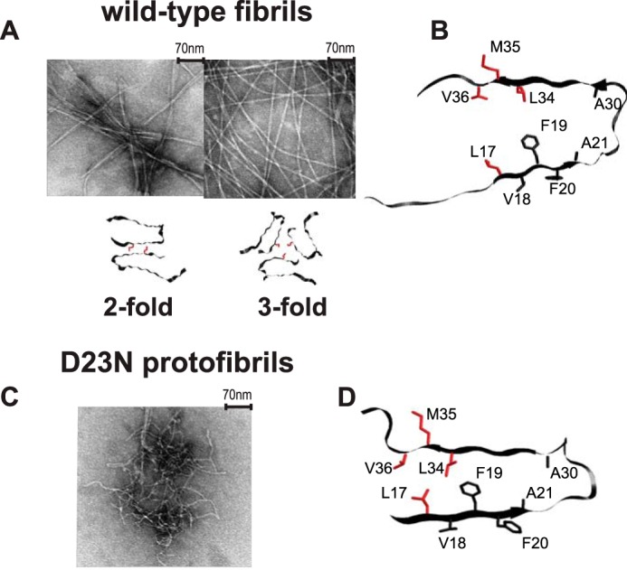
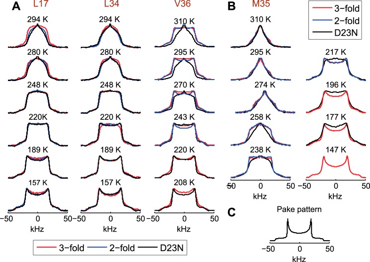
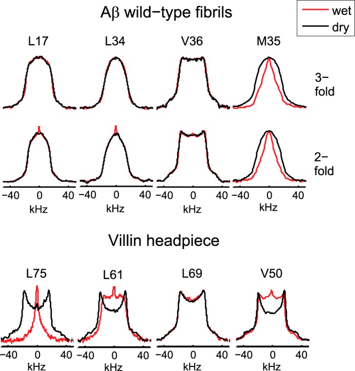
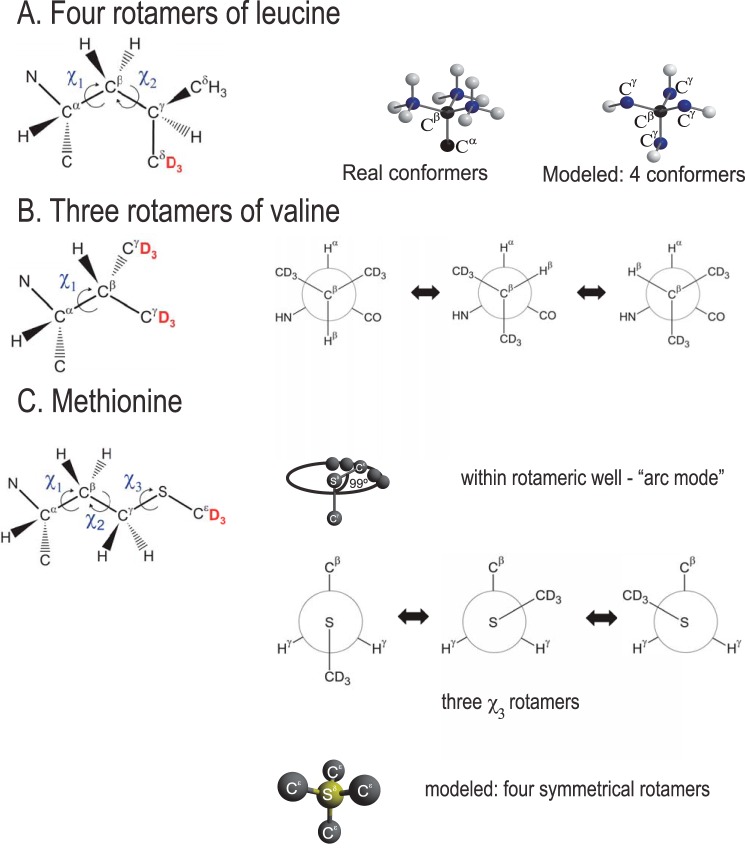
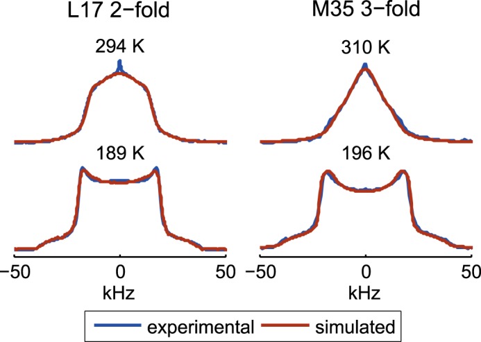
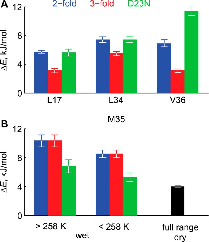
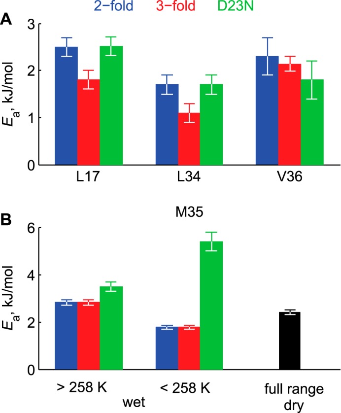
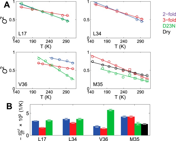
Amyloid fibril deposits found in Alzheimer disease patients are composed of amyloid-β (Aβ) protein forming a number of hydrophobic interfaces that are believed to be mostly rigid. We have investigated the μs-ms time-scale dynamics of the intra-strand hydrophobic core and interfaces of the fibrils composed of Aβ1-40 protein. Using solid-state (2)H NMR line shape experiments performed on selectively deuterated methyl groups, we probed the 3-fold symmetric and 2-fold symmetric polymorphs of native Aβ as well as the protofibrils of D23N Iowa mutant, associated with an early onset of Alzheimer disease. The dynamics of the hydrophobic regions probed at Leu-17, Leu-34, Val-36, and Met-35 side chains were found to be very pronounced at all sites and in all polymorphs of Aβ, with methyl axis motions persisting down to 230-200 K for most of the sites. The dominant mode of motions is the rotameric side chain jumps, with the Met-35 displaying the most complex multi-modal behavior. There are distinct differences in the dynamics among the three protein variants, with the Val-36 site displaying the most variability. Solvation of the fibrils does not affect methyl group motions within the hydrophobic core of individual cross-β subunits but has a clear effect on the motions at the hydrophobic interface between the cross-β subunits, which is defined by Met-35 contacts. In particular, hydration activates transitions between additional rotameric states that are not sampled in the dry protein. Thus, these results support the existence of water-accessible cavity recently predicted by molecular dynamics simulations and suggested by cryo-EM studies.

Keywords: Alzheimer disease; amyloid; amyloid-β (AB); protein dynamic; solid state NMR; solvation.


Figures

FIGURE 1.
FIGURE 1.
A, examples of transmission electron microscopy images of the wild-type fibrils composed of Aβ1–40 and the corresponding quaternary structures in which the position of Met-35 side chains are color-coded in red. The following polymorphs are shown: striated-ribbon/2-fold symmetric polymorph of native Aβ1–40 (PDB ID 2LMN) and twisted/3-fold symmetric polymorph of native Aβ1–40 (PDB ID 2LMP). B, a ribbon diagram of the monomer corresponding to the wild-type Aβ1–40 in the 2-fold morphology with the side chains of Leu-17, Leu-34, Val-36, and Met-35 investigated in this work shown in red. The 3-fold morphology has a very similar monomeric unit. C, an example of transmission electron microscopy image of the D23N mutant Aβ1–40 protofibrils with antiparallel β-sheet structure. D, a ribbon diagram of the D23N protofibrils with the side chains of Leu-17, Leu-34, Val-36, and Met-35 investigated in this work shown in red (PDB ID 2LNQ).
FIGURE 2.
FIGURE 2.
Experimental 2H NMR static line shapes for Leu-17, Leu-34, and Val-36 (A) and Met-35 (B) in the native Aβ1–40 2-fold (blue) and 3-fold (red) symmetric polymorphs and in the D23N mutant protofibrils (black). C, a simulated Pake-pattern for a methyl group undergoing fast methyl rotations in the absence of μs-ms motions. The experimental spectra were collected twice to ensure reproducibility. Additionally, the following replicate samples were prepared and analyzed to ensure reproducibility: Leu-34-labeled in the 2- and 3-fold polymorphs, Leu-17-labeled in the 3-fold polymorph, and Met-35 in the 3-fold polymorph.
FIGURE 3.
FIGURE 3.
Experimental line shapes for the dry (black) and hydrated (red) states. Upper panel, Aβ1–40 wild-type fibrils at 294 K for Leu-17, Leu-34, and Val-36 and at 310 K for Met-35. Lower panel, globular villin headpiece protein hydrophobic core residues at 298 K (56).
FIGURE 4.
FIGURE 4.
A, motional models of leucine. Rotameric jumps around χ1 and χ2 angle are represented by four magnetically non-equivalent conformers (out of nine possible configurations) pointing toward the corners of a tetrahedron. White spheres correspond to the Cδ position, blue spheres correspond to Cγ, and black spheres are for Cβ and Cα, the positions of which are not changed during the rotameric inter-conversions. B, valine. Rotameric jumps around the χ1 angle are represented by three conformers. C, methionine. Arc motion mode (motions within rotameric potential) is used for all states and all temperatures. Rotameric motions mode involving only the χ3 angle is used for the dry state at all temperatures and for the hydrated state at T < 250 K. For the hydrated state at T > 250 K, rotameric motions (involving in reality all three χ1, χ2, and χ3 angles) are approximated by four artificial symmetrical conformers. Positions of the 2H labels at the methyl groups of the side chains are shown in red.
FIGURE 5.
FIGURE 5.
Examples of overlays of the experimental (blue) and fitted spectra (brown line) shown for the Leu-17 site in the 2-fold morphology and the Met-35 site in the 3-fold morphology in the hydrated state.
FIGURE 6.
FIGURE 6.
Energy differences ΔE between the major and minor rotamers. A, hydrophobic core residues. The values for the dry and hydrated samples are identical. B, Met-35 site. The values for the 2-fold and 3-fold polymorphs are identical in both the dry and hydrated states.
FIGURE 7.
FIGURE 7.
Activation energies Ea of rotameric jumps. A, hydrophobic core residues. The values for the dry and hydrated samples are identical. B, Met-35 site. The values for the 2-fold and 3-fold polymorphs are identical in both the dry and hydrated states.
FIGURE 8.
FIGURE 8.
A, the values of the squared order parameters of methyl axes O2 as a function of temperature T. The solid lines represent linear fits. B, the values of dO2/dT.

References

    1. Paravastu A. K., Qahwash I., Leapman R. D., Meredith S. C., and Tycko R. (2009) Seeded growth of β-amyloid fibrils from Alzheimer's brain-derived fibrils produces a distinct fibril structure. Proc. Natl. Acad. Sci. U.S.A. 106, 7443–7448 - PMC - PubMed
    1. Ahmed A. B., and Kajava A. V. (2013) Breaking the amyloidogenicity code: methods to predict amyloids from amino acid sequence. FEBS Lett. 587, 1089–1095 - PubMed
    1. Cannon M. J., Williams A. D., Wetzel R., and Myszka D. G. (2004) Kinetic analysis of β-amyloid fibril elongation. Anal. Biochem. 328, 67–75 - PubMed
    1. Chimon S., Jones C., Calero D. C., and Ishii Y. (2007) A missing link in amyloid misfolding: structural insights into amyloid intermediates for Alzheimer's β-amyloid, Aβ(1–40) by solid-state NMR. Biophys. J. S, 195A–195A
    1. Fitzpatrick A. W., Debelouchina G. T., Bayro M. J., Clare D. K., Caporini M. A., Bajaj V. S., Jaroniec C. P., Wang L., Ladizhansky V., Müller S. A., MacPhee C. E., Waudby C. A., Mott H. R., De Simone A., Knowles T. P. (2013) Atomic structure and hierarchical assembly of a cross-β amyloid fibril. Proc. Natl. Acad. Sci. U.S.A. 110, 5468–5473 - PMC - PubMed

Publication types

MeSH terms

LinkOut - more resources