. 2016 Aug 26;291(35):18453-64.
doi: 10.1074/jbc.M116.729285. Epub 2016 Jul 11.

Protease-activated Receptor-4 Signaling and Trafficking Is Regulated by the Clathrin Adaptor Protein Complex-2 Independent of β-Arrestins

Affiliations

Protease-activated Receptor-4 Signaling and Trafficking Is Regulated by the Clathrin Adaptor Protein Complex-2 Independent of β-Arrestins

Thomas H Smith et al. J Biol Chem. .

Abstract

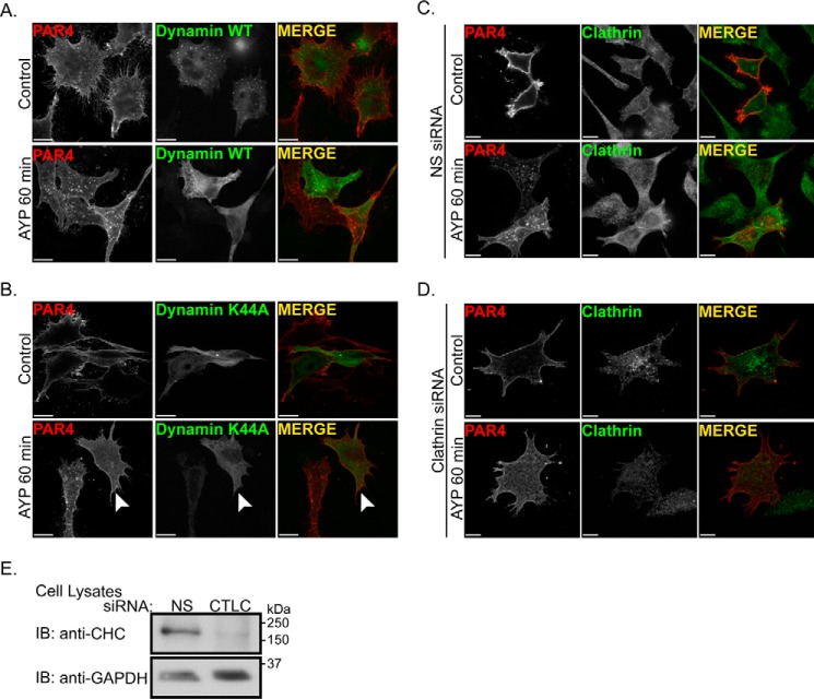
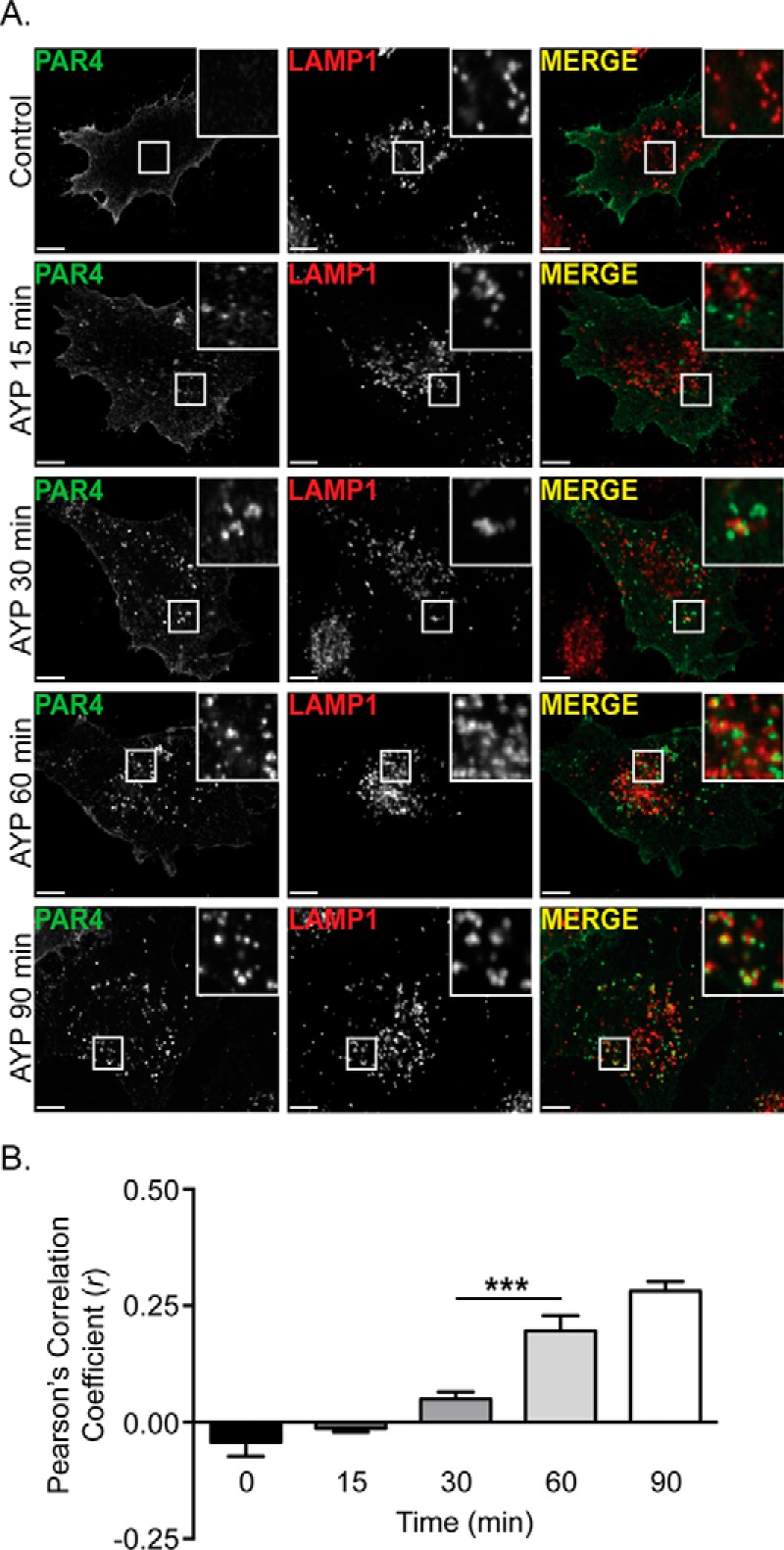
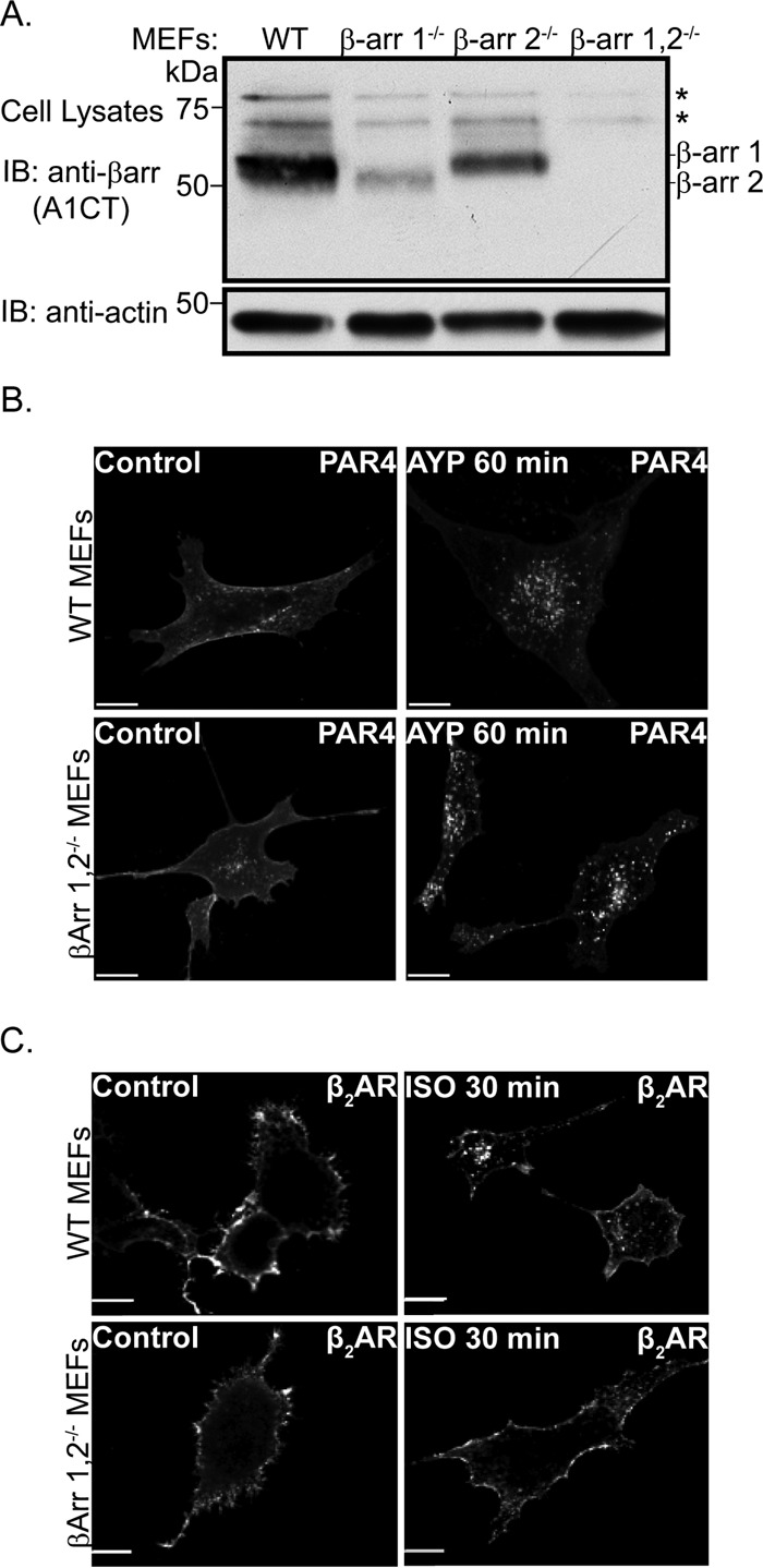
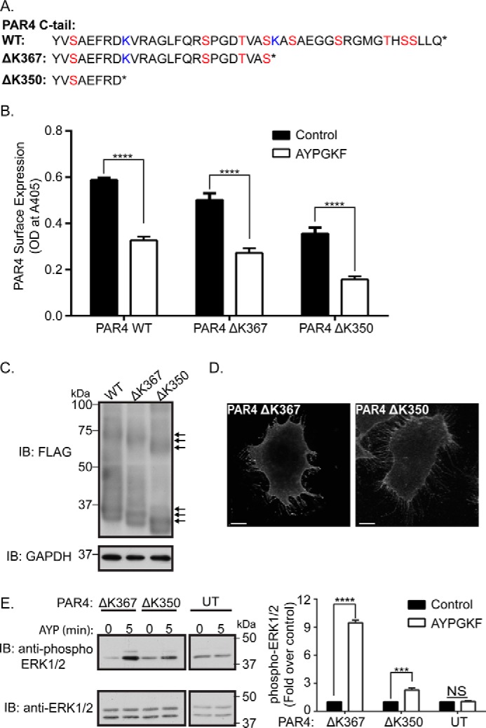
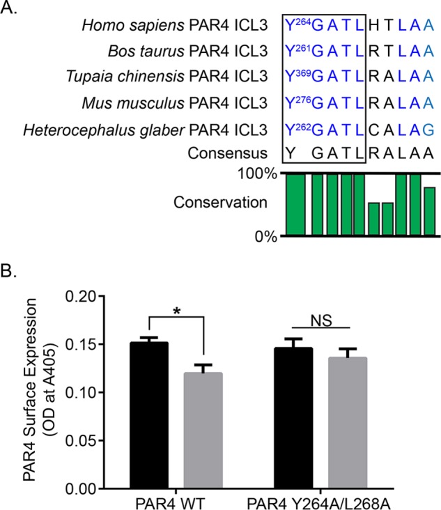
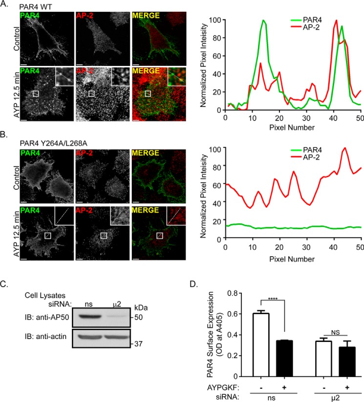
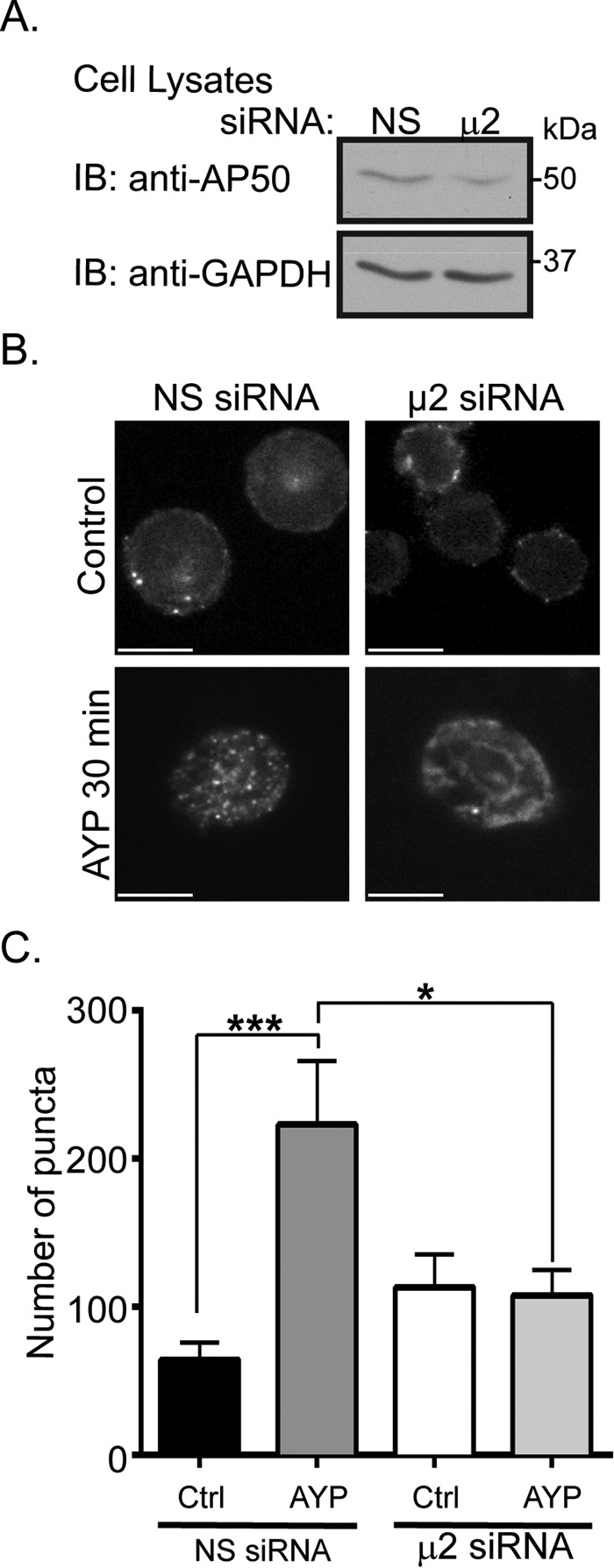
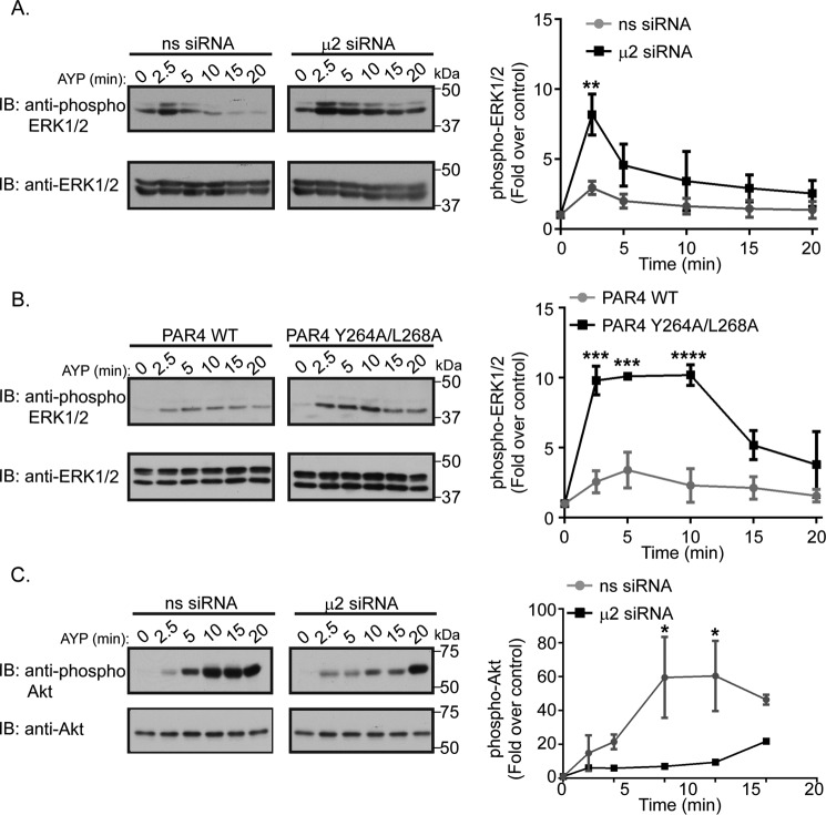
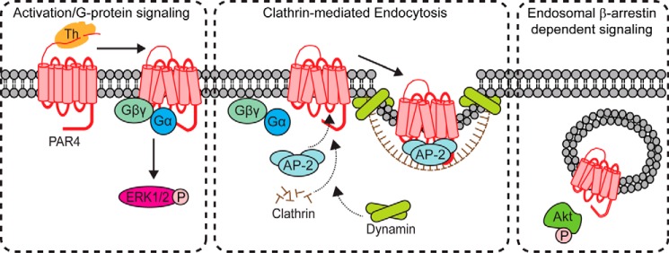
Protease-activated receptor-4 (PAR4) is a G protein-coupled receptor (GPCR) for thrombin and is proteolytically activated, similar to the prototypical PAR1. Due to the irreversible activation of PAR1, receptor trafficking is intimately linked to signal regulation. However, unlike PAR1, the mechanisms that control PAR4 trafficking are not known. Here, we sought to define the mechanisms that control PAR4 trafficking and signaling. In HeLa cells depleted of clathrin by siRNA, activated PAR4 failed to internalize. Consistent with clathrin-mediated endocytosis, expression of a dynamin dominant-negative K44A mutant also blocked activated PAR4 internalization. However, unlike most GPCRs, PAR4 internalization occurred independently of β-arrestins and the receptor's C-tail domain. Rather, we discovered a highly conserved tyrosine-based motif in the third intracellular loop of PAR4 and found that the clathrin adaptor protein complex-2 (AP-2) is important for internalization. Depletion of AP-2 inhibited PAR4 internalization induced by agonist. In addition, mutation of the critical residues of the tyrosine-based motif disrupted agonist-induced PAR4 internalization. Using Dami megakaryocytic cells, we confirmed that AP-2 is required for agonist-induced internalization of endogenous PAR4. Moreover, inhibition of activated PAR4 internalization enhanced ERK1/2 signaling, whereas Akt signaling was markedly diminished. These findings indicate that activated PAR4 internalization requires AP-2 and a tyrosine-based motif and occurs independent of β-arrestins, unlike most classical GPCRs. Moreover, these findings are the first to show that internalization of activated PAR4 is linked to proper ERK1/2 and Akt activation.

Keywords: Akt; G protein-coupled receptor (GPCR); adaptor protein complex-2; arrestin; clathrin; extracellular-signal-regulated kinase (ERK); lysosome; megakaryocyte; thrombin.


Figures

FIGURE 1.
FIGURE 1.
Internalization of activated PAR4 occurs through a dynamin- and clathrin-dependent pathway. A and B, HeLa cells transiently transfected with FLAG-PAR4 and either dynamin WT fused to GFP (A) or the dynamin dominant-negative K44A mutant fused to GFP (B) were pre-labeled with anti-FLAG antibody and stimulated with 500 μm AYPGKF for 60 min at 37 °C. Cells were fixed, permeabilized, immunostained, and imaged by confocal microscopy. Arrowheads, dynamin-GFP K44A mutant expressing cells. The images are representative of three independent experiments (scale bar, 10 μm). C and D, HeLa cells were transiently transfected with FLAG-PAR4 and either nonspecific (NS) or clathrin heavy chain (CHC)-specific siRNA. Cells were incubated with 500 μm AYPGKF for 60 min at 37 °C, processed, and imaged by confocal microscopy. Images are representative of three independent experiments (scale bar, 10 μm). E, cell lysates were collected in parallel, resolved by SDS-PAGE, and immunoblotted (IB) as indicated.
FIGURE 2.
FIGURE 2.
Activated PAR4 is internalized and sorted to early endosomes. A, HeLa cells were transfected with FLAG-PAR4, prelabeled with anti-FLAG antibody, stimulated with 500 μm AYPGKF for various times at 37 °C, processed, co-stained with anti-EEA1 antibody, and imaged by confocal microscopy. The images are representative of three independent experiments (scale bar, 10 μm). Insets, magnifications of boxed areas. B, Pearson's correlation coefficient was determined to quantify the degree of correlation in signal intensity between PAR4 and EEA1 at each pixel. Data (mean ± S.E.; n ≥ 9 cells for each time point) shown were collected from three independent experiments, and statistical significance was determined by one-way ANOVA (*, p < 0.05; ****, p < 0.0001).
FIGURE 3.
FIGURE 3.
Activated internalized PAR4 is sorted to late endosomes/lysosomes. A, HeLa cells transfected with FLAG-PAR4 were prelabeled with anti-FLAG antibody, stimulated with 500 μm AYPGKF for various times at 37 °C, processed, co-stained with anti-LAMP1 antibody, and imaged by confocal microscopy. The images are representative of three independent experiments (scale bar, 10 μm). Insets, magnifications of boxed areas. B, Pearson's correlation coefficient was determined to quantify the degree of correlation in signal intensity between PAR4 and LAMP1 at each pixel. Data (mean ± S.E.; n ≥ 9 cells for each time point) shown were collected from three independent experiments, and statistical significance was determined by one-way ANOVA (***, p < 0.001).
FIGURE 4.
FIGURE 4.
Internalization of activated PAR4 occurs independent of β-arrestins. A, cell lysates from WT and β-arrestin 1/2 double knock-out (β-arr 1,2−/−) MEFs were immunoblotted (IB) as indicated. WT or β-arrestin 1/2 double knock-out MEFs were transiently transfected with FLAG-PAR4 (B) or FLAG-β2AR (C), prelabeled with anti-FLAG antibody, and stimulated with 500 μm AYPGKF for 60 min at 37 °C (B) or 10 nm isoproterenol for 30 min at 37°C (C). After agonist stimulation, cells were processed and imaged by confocal microscopy. The images are representative of several cells from three independent experiments.
FIGURE 5.
FIGURE 5.
The PAR4 C-tail is not required for internalization. A, PAR4 amino acid sequence of full-length receptor and ΔK367 and ΔK350 C-tail truncation mutants. B, HeLa cells transfected with FLAG-PAR4 WT, ΔK367, or ΔK350 mutants were prelabeled with anti-FLAG antibody and stimulated with 500 μm AYPGKF for 60 min at 37 °C. Cell surface ELISA was then used to quantify the amount of PAR4 remaining at the cell surface following stimulation. Data (mean ± S.E.) are representative of three independent experiments, and statistical significance was determined by two-way ANOVA (****, p < 0.0001). OD, optical density. C, cell lysates from HeLa cells transfected as above in B were immunoblotted (IB) as indicated. Arrows, mobility shifts representing differentially processed PAR4 species. D, HeLa cells transfected as above in B were labeled with anti-FLAG antibody, processed, and imaged by confocal microscopy. The images are representative of several cells from three independent experiments. E, untransfected (UT) HeLa cells and HeLa cells transfected as above in B were stimulated with 500 μm AYPGKF (AYP) for 5 min at 37 °C. Cell lysates were resolved by SDS-PAGE and immunoblotted as indicated. Changes in phospho-ERK1/2 signals were quantified, normalized to total ERK1/2, and expressed as a fraction of the untreated control. Samples were resolved on the same gel and separated for labeling. Data (mean ± S.E.) are representative of three independent experiments, and statistical significance was determined by two-way ANOVA (***, p < 0.001; ****, p < 0.0001; NS, not significant).
FIGURE 6.
FIGURE 6.
PAR4 harbors a highly conserved tyrosine-based motif that is important for internalization. A, alignment of human PAR4 intracellular loop 3 (ICL3) sequence with several mammalian orthologues. A highly conserved YX3L motif is indicated by the black box. B, HeLa cells were transfected with FLAG-PAR4 WT or the tyrosine motif Y264A/L268A mutant, prelabeled with anti-FLAG antibody, and stimulated with 500 μm AYPGKF for 60 min at 37 °C. Cell surface ELISA was then used to quantify the amount of PAR4 remaining at the cell surface after stimulation. Data shown (mean ± S.E.) are representative of four independent experiments, and statistical significance was calculated by two-way ANOVA (*, p < 0.05; NS, not significant).
FIGURE 7.
FIGURE 7.
AP-2 mediates internalization of activated PAR4. A and B, HeLa cells transfected with FLAG-PAR4 WT (A) or the Y264A/L268A mutant (B) were prelabeled with anti-FLAG antibody, stimulated with 500 μm AYPGKF for 12.5 min at 37 °C, processed, co-stained with anti-AP-2 antibody, and imaged by confocal microscopy. The images are representative of three independent experiments (scale bar, 10 μm). Insets, magnifications of boxed areas. The fluorescence intensity line scans were generated from the regions denoted by the white dashed line in the agonist-stimulated images (A and B, lower panels). C, HeLa cells expressing FLAG-PAR4 were transfected with nonspecific (ns) siRNA or siRNA specific for the μ2 adaptin subunit of AP-2 and immunoblotted (IB) as indicated. D, HeLa cells transfected as described above in C were prelabeled with anti-FLAG antibody and stimulated with 500 μm AYPGKF for 60 min at 37 °C. ELISA was then used to quantify the amount of receptor at the cell surface. Data shown (mean ± S.E.) are representative of three independent experiments, and statistical significance was calculated by two-way ANOVA (****, p < 0.0001; ns, not significant).
FIGURE 8.
FIGURE 8.
AP-2 is required for internalization of endogenous PAR4 in Dami cells. A, Dami cells were transfected with nonspecific (ns) siRNA or siRNA targeting the μ2 adaptin subunit of AP-2. Cell lysates were collected and immunoblotted (IB) as indicated. B, Dami cells transfected as above in A were prelabeled with anti-PAR4 antibody, treated with 500 μm AYPGKF (AYP) or left untreated (Control) for 30 min at 37 °C, processed, and imaged by confocal microscopy. The images are representative of three independent experiments (scale bar, 10 μm). C, PAR4 internalization was quantified using Slidebook 5.0 software's automated object counting. At least 10 cells were analyzed for each condition for each of three independent experiments. Data shown are combined from three independent experiments and were analyzed using a Student's t test (*, p < 0.05; ***, p < 0.001).
FIGURE 9.
FIGURE 9.
PAR4 trafficking is linked to proper ERK1/2 and Akt signaling. A and C, Dami cells transfected with nonspecific (ns) siRNA or siRNA targeting the μ2 adaptin subunit of AP-2 were stimulated with 500 μm AYPGKF (AYP) for various times at 37 °C. Cell lysates were resolved by SDS-PAGE and immunoblotted (IB) as indicated. Changes in phospho-ERK1/2 (A) and phospho-Akt (C) signals were quantified, normalized to total ERK1/2 and total Akt, respectively, and expressed as a fraction of the untreated controls. B, HeLa cells transfected with FLAG-PAR4 WT or Y264A/L268A mutant were stimulated with 500 μm AYPGKF for various times at 37 °C. Cell lysates were resolved by SDS-PAGE and immunoblotted as indicated. Changes in phospho-ERK1/2 signals were quantified, normalized to total ERK1/2, and expressed as a fraction of the untreated controls. Samples were resolved on the same gel and separated for labeling. Data shown (mean ± S.E.) are representative of three independent experiments, and statistical significance was calculated by two-way ANOVA (*, p < 0.05; **, p < 0.01; ***, p < 0.001; ****, p < 0.0001).
FIGURE 10.
FIGURE 10.
Model of PAR4 trafficking and signaling. PAR4 is a seven transmembrane GPCR that is cleaved and activated by thrombin. Thrombin cleavage generates an N terminus that binds intramolecularly to the receptor, facilitating coupling to heterotrimeric G proteins, which promotes ERK1/2 signaling. After activation, PAR4 is recruited to clathrin-coated pits and requires both an intact tyrosine-based motif and AP-2 for internalization. Once internalized, PAR4 is sorted to early endosomes and appears to stimulate Akt signaling.

References

    1. Coughlin S. R. (2005) Protease-activated receptors in hemostasis, thrombosis, and vascular biology. J. Thromb. Haemost. 3, 1800–1814 - PubMed
    1. Vu T. K., Hung D. T., Wheaton V. I., and Coughlin S. R. (1991) Molecular cloning of a functional thrombin receptor reveals a novel proteolytic mechanism of receptor activation. Cell 64, 1057–1068 - PubMed
    1. Vu T. K., Wheaton V. I., Hung D. T., Charo I., and Coughlin S. R. (1991) Domains specifying thrombin-receptor interaction. Nature 353, 674–677 - PubMed
    1. Scarborough R. M., Naughton M. A., Teng W., Hung D. T., Rose J., Vu T. K., Wheaton V. I., Turck C. W., and Coughlin S. R. (1992) Tethered ligand agonist peptides: structural requirements for thrombin receptor activation reveal mechanism of proteolytic unmasking of agonist function. J. Biol. Chem. 267, 13146–13149 - PubMed
    1. Trejo J., Hammes S. R., and Coughlin S. R. (1998) Termination of signaling by protease-activated receptor-1 is linked to lysosomal sorting. Proc. Natl. Acad. Sci. U.S.A. 95, 13698–13702 - PMC - PubMed