. 2016 Sep 2;291(36):18959-66.
doi: 10.1074/jbc.M116.730176. Epub 2016 Jul 8.

Electrochemical Evidence for Neuroglobin Activity on NO at Physiological Concentrations

Affiliations

Electrochemical Evidence for Neuroglobin Activity on NO at Physiological Concentrations

Stanislav Trashin et al. J Biol Chem. .

Abstract

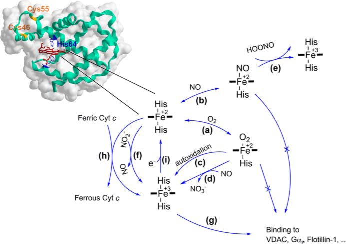
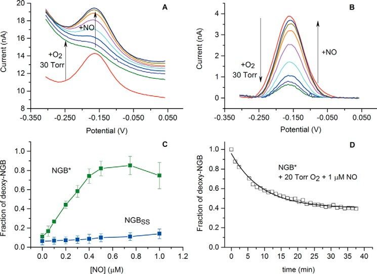
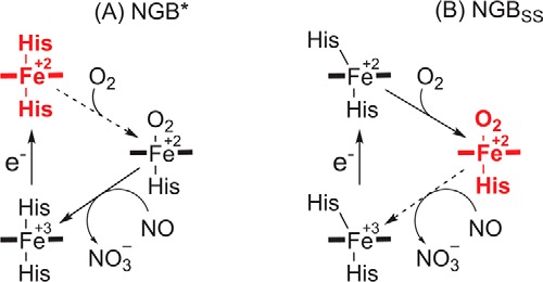
The true function of neuroglobin (Ngb) and, particularly, human Ngb (NGB) has been under debate since its discovery 15 years ago. It has been expected to play a role in oxygen binding/supply, but a variety of other functions have been put forward, including NO dioxygenase activity. However, in vitro studies that could unravel these potential roles have been hampered by the lack of an Ngb-specific reductase. In this work, we used electrochemical measurements to investigate the role of an intermittent internal disulfide bridge in determining NO oxidation kinetics at physiological NO concentrations. The use of a polarized electrode to efficiently interconvert the ferric (Fe(3+)) and ferrous (Fe(2+)) forms of an immobilized NGB showed that the disulfide bridge both defines the kinetics of NO dioxygenase activity and regulates appearance of the free ferrous deoxy-NGB, which is the redox active form of the protein in contrast to oxy-NGB. Our studies further identified a role for the distal histidine, interacting with the hexacoordinated iron atom of the heme, in oxidation kinetics. These findings may be relevant in vivo, for example, in blocking apoptosis by reduction of ferric cytochrome c, and gentle tuning of NO concentration in the tissues.

Keywords: electrochemistry; electron transfer; neuroglobin; nitric oxide; oxygen binding; protein chemistry.


Figures

FIGURE 1.
FIGURE 1.
The relationship of the redox and ligation states of the heme iron and NGB reactivity. a, ferrous (Fe2+) NGB reacts with O2 to form the O2 adduct. The dissociation of the distal histidine limits the binding kinetics due a low dissociation rate constant of 0.6 and 7 s−1 for NGB with reduced and oxidized states of the internal disulfide bridge, respectively (34, 35). The P50 value was reported in the range of 1–8 torr depending on pH, temperature, and the state of the internal disulfide bound (21). b, ferrous NGB reacts with NO leading to the nitrosyl ligated form with Kd ≈ 1 nm and a slow dissociation rate constant koff ≈ 10−4 to 10−3 s−1 (37). c, NGB-O2 adduct is prone to autoxidation with k ≈ 0.17 min−1 (21). Similar to myoglobin and hemoglobin, NGB has been reported as a scavenger of reactive oxygen and nitrogen species (ROS, RNS); d, NGB shows NO dioxygenase activity with kcat ≈ 300 s−1 (37, 38); e, nitrosyl ferrous NGB but not ferric (Fe3+) NGB can scavenge peroxynitrite with kcat ≈ 1.3 × 10⁵ m−1 s−1 (39); f, ferrous (Fe2+) NGB can reduce nitrite (NO2) although with a relatively low rate constant of 0.06 and 0.12 m−1 s−1 for NGB with the reduced and oxidized states of the disulfide bridge, respectively (40). The role of NGB in blocking apoptosis through binding other proteins has been also suggested in the literature: g, NGB can bind to the GDP-bound form of the α-subunit of heterotrimeric G-protein (Gαi) with Kd ≈ 6 × 10² nm (41). Immunoprecipitation techniques revealed binding of NGB to voltage-dependent anion channel (42), Flotillin-1 (43), two members of the Rho GTPase family (Rac1 and RhoA), as well as the Pak1 kinase (44), and a subunit of the mitochondrial complex III cytochrome c1 (45). It has been found that the conformational transition in NGB upon binding ligands such as O2 and NO can prevent NGB binding to Gαi. Similar mechanisms may regulate binding to other proteins. h, moreover, ferrous NGB is capable to reduce rapidly ferric cytochrome c (Cyt c) with a kinetic constant of 2 × 10⁷ m−1 s−1 (46), which seems to be facilitated by an appropriate docking between two proteins (47, 48). Interaction between Cyt c and NGB goes through a rapid transient binding with a relatively high dissociation constant, Kd ≈ 120 μm (48), which decreases in low ionic strength buffer suggesting that the interaction is largely electrostatic in nature (48, 49). i, the reaction that is responsible for one-electron reduction of the ferric (Fe3+) to ferrous (Fe2+) form is not known yet. It can be driven by an enzyme process or a low molecular weight redox compound/mediator. Noteworthy, hexacoordination of the heme iron favors reduction kinetics comparing to pentacoordinated analogues (50).
FIGURE 2.
FIGURE 2.
CV and DPV behavior of NGB* in PBS buffer solution (A and B) and immobilized on the gold electrodes (C and D). Concentration of the dissolved NGB*, 20 μm; surface density of the immobilized NGB*, 2 pmol/cm2; CV scan rate, 20 mV/s; DPV modulation amplitude, 20 mV; modulation time, 0.05 s; interval time, 0.5 s. The arrows denote scan directions. Blank voltammograms are shown by dashed lines (in green) and curves recorded in the air-saturated buffer are presented by dashed-dotted lines (in blue).
SCHEME 1
SCHEME 1
FIGURE 3.
FIGURE 3.
Behavior of NGB in the presence of different levels of O2. A and B, change in voltammograms with an increase of O2 level in the presence of NGB* and NGBSS; C, determined fraction of the oxygenated NGB in equilibrium with the ferrous (Fe+2) NGB as a function of O2 concentration. Error bars show standard deviation for three consecutive measurements. PBS, pH 7.4, 25 °C.
FIGURE 4.
FIGURE 4.
Behavior of NGB in the presence of 30 torr O2 and submicromolar NO concentrations. A, change in DPV curves for NGB*; B, the DPV curves after baseline correction; C, dependence of the steady state deoxy-NGB fraction on NO concentration in the presence of 30 torr O2. Average values of three separate titration curves are plotted, with standard deviation values. D, stability of deoxy-NGB* in the presence of 20 torr O2 and 1 μm NO. Arrows denote direction of changes. PBS, pH 7.4, 25 °C.
FIGURE 5.
FIGURE 5.
The mechanism of NGB transitions in the presence of 0.5 μm NO and 30 torr O2. A, behavior of NGB*, which models the protein with the opened disulfide bridge that favors the hexacoordinate ferrous form; B, behavior of NGBSS, which is the protein with the disulfide bridge that favors the oxygenated form. Broken arrows indicate a rate-limiting step and the bold red structures depict an accumulated form.

References

    1. Burmester T., Weich B., Reinhardt S., and Hankeln T. (2000) A vertebrate globin expressed in the brain. Nature 407, 520–523 - PubMed
    1. Tejero J., and Gladwin M. T. (2014) The globin superfamily: functions in nitric oxide formation and decay. Biol. Chem. 395, 631–639 - PMC - PubMed
    1. Burmester T., and Hankeln T. (2014) Function and evolution of vertebrate globins. Acta Physiol. 211, 501–514 - PubMed
    1. Ascenzi P., Gustincich S., and Marino M. (2014) Mammalian nerve globins in search of functions. IUBMB Life 66, 268–276 - PubMed
    1. Haines B., Demaria M., Mao X., Xie L., Campisi J., Jin K., and Greenberg D. A. (2012) Hypoxia-inducible factor-1 and neuroglobin expression. Neurosci. Lett. 514, 137–140 - PMC - PubMed

LinkOut - more resources