

Imaging assessment of glenohumeral dysplasia secondary to brachial plexus birth palsy
- PMID: 27403013
- PMCID: PMC4938443
- DOI: 10.1590/0100-3984.2015.0039
Imaging assessment of glenohumeral dysplasia secondary to brachial plexus birth palsy
Abstract
Objective: To assess imaging parameters related to the morphology of the glenohumeral joint in children with unilateral brachial plexus birth palsy (BPBP), in comparison with those obtained for healthy shoulders.
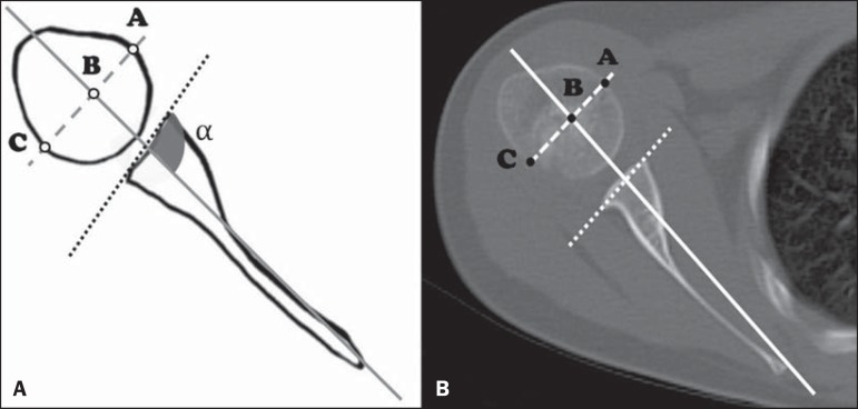
Materials and methods: We conducted a retrospective search for cases of unilateral BPBP diagnosed at our facility. Only patients with a clinical diagnosis of unilateral BPBP were included, and the final study sample consisted of 10 consecutive patients who were assessed with cross-sectional imaging. The glenoid version, the translation of the humeral head, and the degrees of glenohumeral dysplasia were assessed.
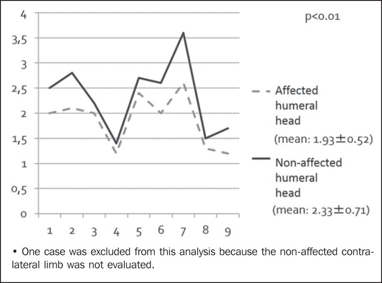
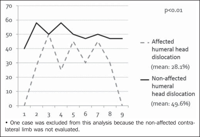
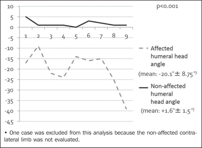
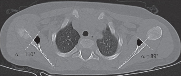
Results: The mean diameter of the affected humeral heads was 1.93 cm, compared with 2.33 cm for those of the normal limbs. In two cases, there was no significant posterior displacement of the humeral head, five cases showed posterior subluxation of the humeral head, and the remaining three cases showed total luxation of the humeral head. The mean glenoid version angle of the affected limbs (90-α) was -9.6º, versus +1.6º for the normal, contralateral limbs.
Conclusion: The main deformities found in this study were BPBP-associated retroversion of the glenoid cavity, developmental delay of the humeral head, and posterior translation of the humeral head.
Objetivo: Avaliar os parâmetros de imagem relacionados com a morfologia da articulação glenoumeral em crianças com paralisia obstétrica do plexo braquial (POPB) unilateral, comparando-os com os observados em ombros saudáveis.
Materiais e métodos: Foi realizada uma busca retrospectiva de casos de POPB unilateral diagnosticados em nossa instituição. Somente foram incluídos os pacientes com diagnóstico clínico de POPB unilateral, e a amostra final do estudo consistiu em 10 pacientes consecutivos avaliados por meio de imagens transversais. Foram avaliados a retroversão da cavidade glenoide, a translação da cabeça do úmero e o grau de displasia glenoumeral.
Resultados: A média do diâmetro da cabeça do úmero foi 1,93 cm nos membros afetados e 2,33 cm nos membros normais. Em dois casos, não houve deslocamento posterior significativo da cabeça do úmero, cinco casos apresentaram subluxação posterior da cabeça do úmero, e os três casos restantes apresentaram luxação total da cabeça do úmero. A média do ângulo de retroversão glenoide dos membros afetados (90-α) foi -9,6º, ao passo que a dos membros contralaterais normais foi +1,6º.
Conclusão: As principais deformidades encontradas neste estudo foram retroversão da cavidade glenoide relacionada com POPB, atraso no desenvolvimento da cabeça do úmero e translação posterior da cabeça do úmero.
Keywords: Birth injuries/complications; Brachial plexus neuropathies/complications; Humeral head/abnormalities; Joint diseases/diagnosis; Shoulder dislocation/diagnosis; Tomography.
Figures
Similar articles
-
Quantification of humeral head deformity following neonatal brachial plexus palsy.J Bone Joint Surg Am. 2012 Sep 19;94(18):e136(1-8). doi: 10.2106/JBJS.K.00540. J Bone Joint Surg Am. 2012. PMID: 22992884
-
Isolated open anterior shoulder release in brachial plexus birth palsy.J Shoulder Elbow Surg. 2019 Jul;28(7):1347-1355. doi: 10.1016/j.jse.2018.12.016. Epub 2019 Apr 10. J Shoulder Elbow Surg. 2019. PMID: 30981548
-
Microcomputed tomography characterization of shoulder osseous deformity after brachial plexus birth palsy: a rat model study.J Bone Joint Surg Am. 2010 Nov 3;92(15):2583-8. doi: 10.2106/JBJS.I.01660. J Bone Joint Surg Am. 2010. PMID: 21048177
-
External rotation humeral osteotomy for brachial plexus birth palsy.Tech Hand Up Extrem Surg. 2007 Mar;11(1):8-14. doi: 10.1097/01.bth.0000248359.14448.e6. Tech Hand Up Extrem Surg. 2007. PMID: 17536517 Review.
-
Glenohumeral deformity in children with brachial plexus birth injuries.Bull NYU Hosp Jt Dis. 2011;69(1):36-43. Bull NYU Hosp Jt Dis. 2011. PMID: 21332437 Review.
Cited by
-
Modified Friedman technique: a new proposed method of measuring glenoid version in the setting of glenohumeral dysplasia.Pediatr Radiol. 2018 Nov;48(12):1779-1785. doi: 10.1007/s00247-018-4196-7. Epub 2018 Jul 5. Pediatr Radiol. 2018. PMID: 29978295
-
Usefulness of dynamic contrast-enhanced MRI in the evaluation of osteonecrosis of the proximal fragment in scaphoid fractures.Radiol Bras. 2018 Sep-Oct;51(5):334. doi: 10.1590/0100-3984.2017.0036. Radiol Bras. 2018. PMID: 30369662 Free PMC article. No abstract available.
-
Adding Value to the Magnetic Resonance Examination in a Case of Brachial Plexus Birth Palsy.J Clin Imaging Sci. 2018 Aug 24;8:38. doi: 10.4103/jcis.JCIS_26_18. eCollection 2018. J Clin Imaging Sci. 2018. PMID: 30197829 Free PMC article.
-
The Pathogenesis of Glenohumeral Deformity and Contracture Formation in Obstetric Brachial Plexus Palsy-A Review.J Brachial Plex Peripher Nerve Inj. 2019 Jul 12;14(1):e24-e34. doi: 10.1055/s-0039-1692420. eCollection 2019 Jan. J Brachial Plex Peripher Nerve Inj. 2019. PMID: 31308856 Free PMC article. Review.
-
Ischiofemoral impingement secondary to valgus intertrochanteric osteotomy: a case report.Radiol Bras. 2017 Sep-Oct;50(5):335-337. doi: 10.1590/0100-3984.2013.0026. Radiol Bras. 2017. PMID: 29085168 Free PMC article.
References
-
- Ruchelsman DE, Grossman JA, Price AE. Glenohumeral deformity in children with brachial plexus birth injuries. Bull NYU Hosp Jt Dis. 2011;69:36–43. - PubMed
-
- van der Sluijs JA, van Ouwerkerk WJ, de Gast A, et al. Deformities of the shoulder in infants younger than 12 months with an obstetric lesion of the brachial plexus. J Bone Joint Surg Br. 2001;83:551–555. - PubMed
-
- Hoenecke HR, Jr, Hermida JC, Flores-Hernandez C, et al. Accuracy of CT-based measurements of glenoid version for total shoulder arthroplasty. J Shoulder Elbow Surg. 2010;19:166–166. - PubMed
-
- Churchill RS, Brems JJ, Kotschi H. Glenoid size, inclination, and version: an anatomic study. J Shoulder Elbow Surg. 2001;10:327–332. - PubMed
LinkOut - more resources
Full Text Sources
Other Literature Sources