. 2016:2016:5308170.
doi: 10.1155/2016/5308170. Epub 2016 Jun 14.

NF-κB-Regulated miR-99a Modulates Endothelial Cell Inflammation

Affiliations

NF-κB-Regulated miR-99a Modulates Endothelial Cell Inflammation

Mei-Hua Bao et al. Mediators Inflamm. 2016.

Abstract

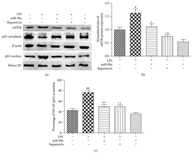
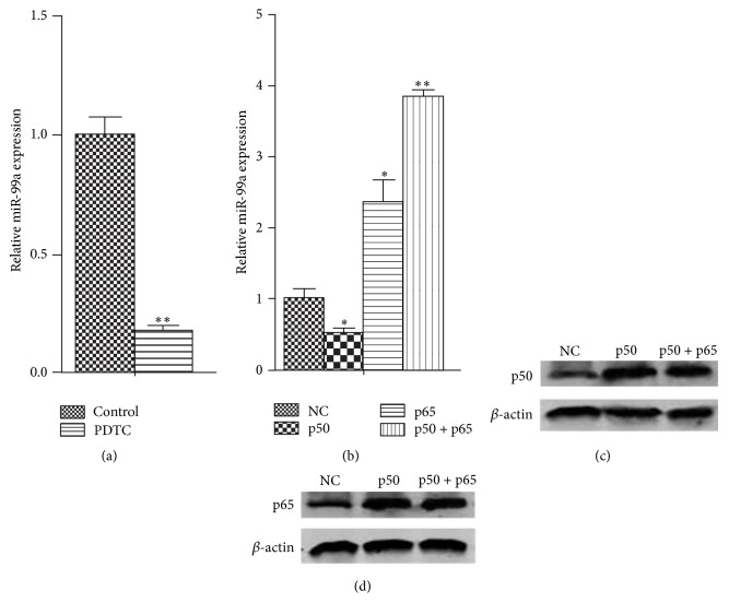
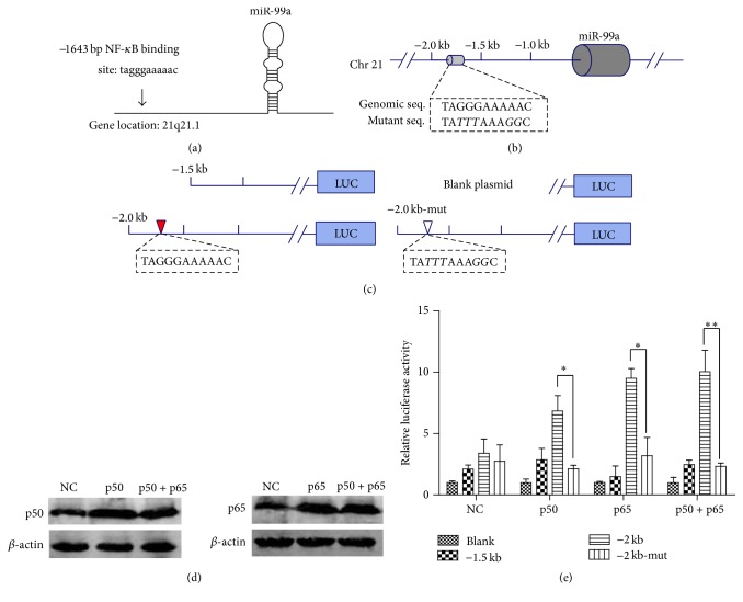
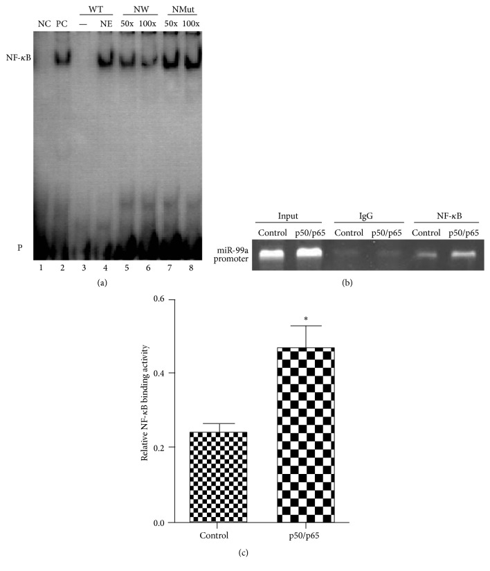
Objective. The present study was performed to investigate the effects and mechanisms of miR-99a on LPS-induced endothelial cell inflammation, as well as the regulation of NF-κB on miR-99a production. Methods and Results. ELISA showed that LPS treatment significantly promoted the secretion of inflammatory factors (TNF-α, IL-6, IL-1β, and MCP-1). LPS treatment also inhibited miR-99a production and promoted mTOR expression and NF-κB nuclear translocation. Overexpression of miR-99a suppressed the LPS-induced TNF-α, IL-6, IL-1β, and MCP-1 overproduction, mTOR upregulation, and NF-κB nuclear translocation. The PROMO software analysis indicated NF-κB binding site in the -1643 to -1652 region of miR-99a promoter. Dual luciferase reporter analysis, electrophoretic mobility shift assays (EMSA), and chromosome immunoprecipitation (ChIP) assays demonstrated that NF-κB promoted the transcription of miR-99a by binding to the -1643 to -1652 region of miR-99a promoter. Further studies on HUVECs verified the regulatory effects of NF-κB on miR-99a production. Conclusion. MiR-99a inhibited the LPS-induced HUVECs inflammation via inhibition of the mTOR/NF-κB signal. NF-κB promoted miR-99a production by binding to the -1643 to -1652 region of miR-99a promoter. Considering the importance of endothelial inflammation on cardiovascular diseases, such as atherosclerosis, our results may provide a new insight into the pathogenesis and therapy of atherosclerosis.


Figures

Figure 1
Figure 1
Effects of LPS on miR-99a production and effects of miR-99a on LPS-induced inflammation in HUVECs. (a) Effects of LPS treatment at different concentrations for 24 hours on miR-99a expression; (b) effects of different time treatment with LPS (20 μg/mL) on miR-99a expression; (c) effects of miR-99a on LPS-induced inflammatory factors secretion. The values (mean ± SD from three independent experiments). # P < 0.05; ## P < 0.01 versus control; P < 0.05; ∗∗ P < 0.01 versus LPS group; ∗∗∗ P < 0.001 versus LPS group.
Figure 2
Figure 2
Effects of miR-99a on LPS-induced mTOR expression and NF-κB nuclear translocation. Endothelial cells were transfected with miR-99a (100 nM) mimic or rapamycin (50 nM) for 24 hours and then treated with LPS (20 μg/mL) for another 24 hours. The protein levels were detected by Western-blot. (a) The expression of different proteins analyzed by Western-blot; (b) quantified band density for mTOR relative to control, which was set as 1; (c) percentage of NF-κB in nucleus, total NF-κB presented by the summary of quantified band density for NF-κB (cytoplasm) and NF-κB (nucleus). All values are presented as mean ± SD from three independent experiments. # P < 0.05; ## P < 0.01 versus control; P < 0.05; ∗∗ P < 0.01 versus LPS.
Figure 3
Figure 3
Regulatory effects of NF-κB on miR-99a expression in HUVECs. ((a), (b)) Relative miR-99a expression in HUVECs after NF-κB overexpression or inhibition. ((c), (d)) The increased p50 or p65 protein levels in HUVECs which were transfected with 4 μg of NF-κB subunit ORF cDNA Clones (p50, p65, or p50 + p65) in 6-well plate for 48 hours. The values (mean ± SD from three independent experiments) are relative to NC or control, which was set as 1. P < 0.05; ∗∗ P < 0.01 versus NC group.
Figure 4
Figure 4
Dual luciferase reporter analysis of the regulate effects of NF-κB on miR-99a promoter plasmids. (a) The predicted NF-κB binding sites in miR-99a promoter analyzed by PROMO software. ((b), (c)) Portions of the upstream region of miR-99a cloned to create −1.5 kb, −2.0 kb, and −2.0 kb-Mut plasmids. (d) The increased p65 or p50 protein levels in HEK293 cells after being transfected with 150 ng of NF-κB subunit ORF cDNA Clones (p50, p65, or p50 + p65) in 24-well plates for 48 hours. (e) Relative luciferase activity of different promoter reporter plasmids after NF-κB overexpression. The values (mean ± SD from three independent experiments) are relative to NC-Blank, which was set as 1. P < 0.05; ∗∗ P < 0.01.
Figure 5
Figure 5
EMSA and ChIP verification of interactions of NF-κB with its binding sites in the miR-99a promoter. (a) EMSA validation of NF-κB binding sites on miR-99a promoter. Lane 1, labeled positive NF-κB probe only; lane 2, nuclear extracts plus labeled positive NF-κB probe; lane 3, labeled WT probe only; lane 4, nuclear extracts plus labeled WT probe; lanes 5 and 6, nuclear extracts plus labeled WT probe and cold unlabeled WT probe (50-fold and 100-fold labeled WT probe), respectively; lanes 7 and 8, nuclear extracts plus labeled WT probe and cold unlabeled Mut probe (50-fold and 100-fold labeled WT probe), respectively. NC: negative control; PC: positive control; NE: nuclear extracts; WT: wild-type probe; NWT: nonlabeled wild-type probe; NMut: nonlabeled mutant probe; P: free biotin-labeled probe. (b) Representative gel images of ChIP assay. (c) Relative NF-κB binding activity quantified by the ChIP bands. P < 0.05 versus control group.

References

    1. Widlansky M. E., Gokce N., Keaney J. F., Jr., Vita J. A. The clinical implications of endothelial dysfunction. Journal of the American College of Cardiology. 2003;42(7):1149–1160. doi: 10.1016/S0735-1097(03)00994-X. - DOI - PubMed
    1. Park K.-H., Park W. J. Endothelial dysfunction: clinical implications in cardiovascular disease and therapeutic approaches. Journal of Korean Medical Science. 2015;30(9):1213–1225. doi: 10.3346/jkms.2015.30.9.1213. - DOI - PMC - PubMed
    1. Tao Z., Zhao H., Wang R., et al. Neuroprotective effect of microRNA-99a against focal cerebral ischemia-reperfusion injury in mice. Journal of the Neurological Sciences. 2015;355(1-2):113–119. doi: 10.1016/j.jns.2015.05.036. - DOI - PubMed
    1. Yu S.-H., Zhang C.-L., Dong F.-S., Zhang Y.-M. miR-99a suppresses the metastasis of human non-small cell lung cancer cells by targeting AKT1 signaling pathway. Journal of Cellular Biochemistry. 2015;116(2):268–276. doi: 10.1002/jcb.24965. - DOI - PubMed
    1. Warth S. C., Hoefig K. P., Hiekel A., et al. Induced miR-99a expression represses Mtor cooperatively with miR-150 to promote regulatory T-cell differentiation. The EMBO Journal. 2015;34(9):1195–1213. doi: 10.15252/embj.201489589. - DOI - PMC - PubMed

MeSH terms