. 2016:2016:8102478.
doi: 10.1155/2016/8102478. Epub 2016 Jun 14.

Therapeutic Potential of Dental Pulp Stem Cell Secretome for Alzheimer's Disease Treatment: An In Vitro Study

Affiliations

Therapeutic Potential of Dental Pulp Stem Cell Secretome for Alzheimer's Disease Treatment: An In Vitro Study

Nermeen El-Moataz Bellah Ahmed et al. Stem Cells Int. 2016.

Abstract

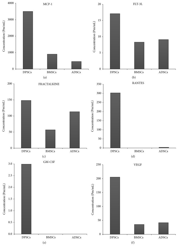
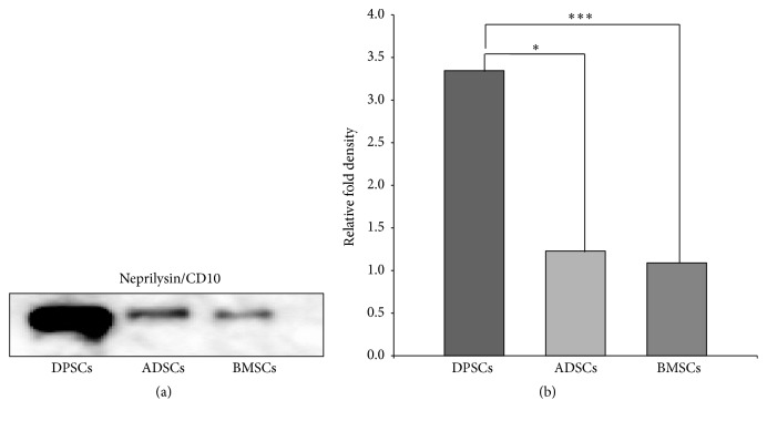
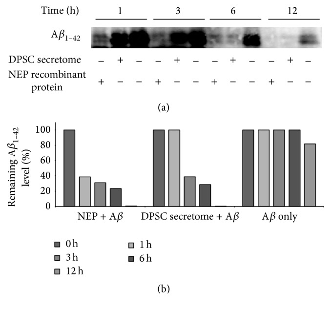
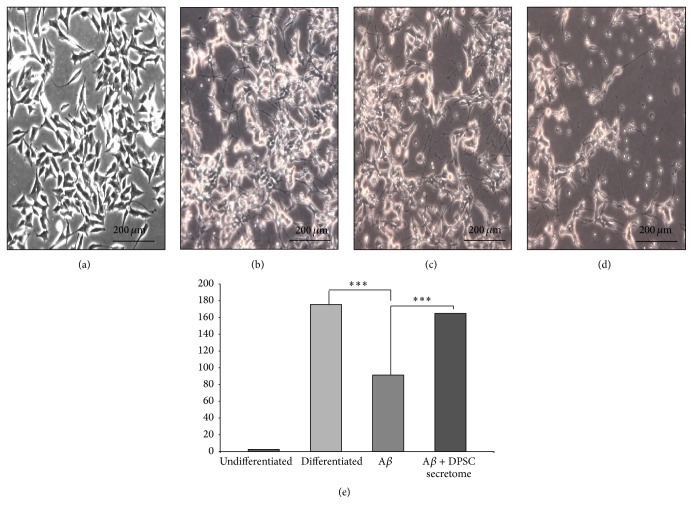
The secretome obtained from stem cell cultures contains an array of neurotrophic factors and cytokines that might have the potential to treat neurodegenerative conditions. Alzheimer's disease (AD) is one of the most common human late onset and sporadic neurodegenerative disorders. Here, we investigated the therapeutic potential of secretome derived from dental pulp stem cells (DPSCs) to reduce cytotoxicity and apoptosis caused by amyloid beta (Aβ) peptide. We determined whether DPSCs can secrete the Aβ-degrading enzyme, neprilysin (NEP), and evaluated the effects of NEP expression in vitro by quantitating Aβ-degrading activity. The results showed that DPSC secretome contains higher concentrations of VEGF, Fractalkine, RANTES, MCP-1, and GM-CSF compared to those of bone marrow and adipose stem cells. Moreover, treatment with DPSC secretome significantly decreased the cytotoxicity of Aβ peptide by increasing cell viability compared to nontreated cells. In addition, DPSC secretome stimulated the endogenous survival factor Bcl-2 and decreased the apoptotic regulator Bax. Furthermore, neprilysin enzyme was detected in DPSC secretome and succeeded in degrading Aβ 1-42 in vitro in 12 hours. In conclusion, our study demonstrates that DPSCs may serve as a promising source for secretome-based treatment of Alzheimer's disease.


Figures

Figure 1
Figure 1
DPSCs secrete some cytokines and growth factors more than both BMSCs and ADSCs. (a) MCP-1. (b) FLT-3L. (c) FRACTALKIN. (d) RANTES. (e) GM-CSF. (f) VEGF.
Figure 2
Figure 2
Exposure to Aβ 1–42 decreased the viability of SH-SY5Y cells in a dose- and time-dependent manner. (a) Phase contrast micrographs of SH-SY5Y cells treated with various concentrations of Aβ 1–42 for different periods of time. (b) Cell viability as detected at 450 nm absorbance (mean ± SE).
Figure 3
Figure 3
DPSC secretome treatment preserves morphology and improves the viability of SH-SY5Y cells exposed to Aβ 1–42. (a–c) Phase contrast micrographs of SH-SY5Y cells exposed to Aβ 1–42 only, Aβ 1–42, and DPSC secretome or nonexposed as control (full-size images are presented in Supplementary Figure S1). (d) Cell viability as detected at 450 nm absorbance (mean ± SE, n = 3. p < 0.05, ∗∗ p < 0.01).
Figure 4
Figure 4
DPSC secretome stimulates the endogenous survival factor Bcl-2 and decreases the apoptotic regulator Bax. (a) Immunoblotting analysis with an anti-Bax, an anti-Bcl2, or an anti-actin antibody (full-length blots are presented in Supplementary Figure S2). (b) Relative fold density as analyzed by Image J (mean ± SE, n = 3. ∗∗ p < 0.01, ∗∗∗ p < 0.001).
Figure 5
Figure 5
DPSC secretome contains a higher concentration of neprilysin/CD10. (a) Immunoblotting analysis with an anti-NEP antibody (full-length blots are presented in Supplementary Figure S3). (b) Relative fold density as analyzed by Image J (mean ± SE, n = 3. p < 0.05, ∗∗∗ p < 0.001).
Figure 6
Figure 6
DPSC secretome degrades Aβ 1–42 protein in vitro. (a) Immunoblotting analysis at each time point with an anti-Aβ antibody (full-length blots are presented in Supplementary Figure S4). (b) Quantitation of remaining Aβ 1–42 level (mean ± SE).
Figure 7
Figure 7
DPSC secretome has neuroprotective ability against Aβ 1–42 induced neurotoxicity. (a–d) Representative photos demonstrating the morphology of SH-SY5Y cells in different treatment groups: undifferentiated, nonexposed differentiated, differentiated exposed to Aβ 1–42 and DPSC secretome, and differentiated exposed to Aβ 1–42 only (full-size images are presented in Supplementary Figure S5) (e). Quantitative analysis of SH-SY5Y neurite outgrowth after exposure to Aβ 1–42 for 24 hours (mean ± SE, n = 3, ∗∗∗ p < 0.001).

Similar articles

Cited by

References

    1. Li M., Guo K., Ikehara S. Stem cell treatment for Alzheimer’s disease. International Journal of Molecular Sciences. 2014;15(10):19226–19238. doi: 10.3390/ijms151019226. - DOI - PMC - PubMed
    1. Alzheimer's Association. Alzheimer's disease facts and figures. Alzheimer's & Dementia. 2013;9(2):208–245. doi: 10.1016/j.jalz.2013.02.003. - DOI - PubMed
    1. Carret-Rebillat A.-S., Pace C., Gourmaud S., et al. Neuroinflammation and Aβ accumulation linked to systemic inflammation are decreased by genetic PKR down-regulation. Scientific Reports. 2015;5, article 8489 doi: 10.1038/srep08489. - DOI - PMC - PubMed
    1. Piermartiri T. C. B., Figueiredo C. P., Rial D., et al. Atorvastatin prevents hippocampal cell death, neuroinflammation and oxidative stress following amyloid-β 1 − 40 administration in mice: evidence for dissociation between cognitive deficits and neuronal damage. Experimental Neurology. 2010;226(2):274–284. doi: 10.1016/j.expneurol.2010.08.030. - DOI - PubMed
    1. Zhou W.-W., Lu S., Su Y.-J., et al. Decreasing oxidative stress and neuroinflammation with a multifunctional peptide rescues memory deficits in mice with Alzheimer disease. Free Radical Biology and Medicine. 2014;74:50–63. doi: 10.1016/j.freeradbiomed.2014.06.013. - DOI - PubMed

LinkOut - more resources