

A Review and Report of Peripheral Giant Cell Granuloma in a 4-Year-Old Child
- PMID: 27403351
- PMCID: PMC4925949
- DOI: 10.1155/2016/7536304
A Review and Report of Peripheral Giant Cell Granuloma in a 4-Year-Old Child
Abstract
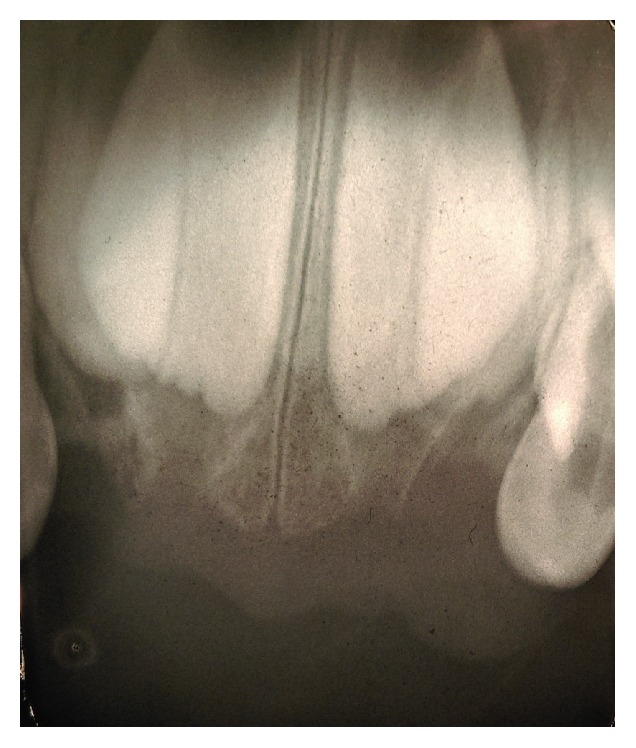
Peripheral giant cell granuloma is a common benign and reactive gingival epulis in oral cavity. It is often difficult to make a clinical diagnosis; thereby definitive diagnosis depends on histopathologic features. We report a case of a 4-year-old Caucasian boy presenting with a five-month history a 20 × 15 × 12 mm pedunculated, lobular soft tissue mass of the left anterior maxilla gingiva which was misdiagnosed and maltreated before his referral. An excisional biopsy of the lesion followed by histopathologic examination of the biopsy specimen revealed distinctive features of peripheral giant cell granuloma. Early detection and excision of this hyperplastic nodule especially in children are important to minimize potential dentoalveolar complications.
Figures


References
-
- Neville B. W., Damm D. D., Allen C. M., Bouquot J. E. Soft tissue tumors. In: Neville B. W., Damm D. D., Allen C. M., Bouquot J. E., editors. Oral and Maxillofacial Pathology. 4th. Philadelphia, Pa, USA: WB Saunders; 2015. p. p. 485.
-
- Flaitz C. M. Peripheral giant cell granuloma: a potentially aggressive lesion in children. Pediatric Dentistry. 2000;22(3):232–233. - PubMed
-
- Soames J. V., Southam J. C. Hyperplastic, neoplastic and related disorders of oral mucosa. In: Soames J. V., editor. Textbook of Oral Pathology. 4th. Oxford, UK: Oxford Medical Publications; 2000. p. p. 103.
LinkOut - more resources
Full Text Sources
Other Literature Sources
