. 2016:2016:6353471.
doi: 10.1155/2016/6353471. Epub 2016 Jun 14.

Arteriovenous Fistula Embolization in Suspected Parauterine Choriocarcinoma

Affiliations

Arteriovenous Fistula Embolization in Suspected Parauterine Choriocarcinoma

Husain Alturkistani et al. Case Rep Obstet Gynecol. 2016.

Abstract

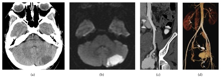
This is a case of choriocarcinoma that did not regress after chemotherapy treatment. A 30-year-old female patient (gravida 2, para 2), presented to our ER with stroke and persistent mild pelvic pain 2 months after a Caesarean section. Computed tomography (CT) revealed an ischemic left hemicerebellar region and a hypervascular mass in the pelvic region. This mass was not present on routine fetal ultrasound during pregnancy. The lesion was treated by chemotherapy after closure of a foramen ovale and insertion of an inferior vena cava (IVC) filter. After that, 2 courses of EMACO (Etoposide, Methotrexate, Actinomycin D, Cyclophosphamide, and Vincristine) chemotherapy regimen were given. Posttreatment CT showed the hypervascular mass without any changes. Arteriography showed the arteriovenous fistulae that were embolized successfully with plugs, coils, and glue. Embolization was considered due to the risk of acute hemorrhagic life-threatening complications. Eight chemotherapy courses were added after embolization. Treatment by endovascular approach and reduction of the hypervascular mass can be a valuable adjunct to chemotherapy treatment of choriocarcinoma.


Figures

Figure 1
Figure 1
(a) Noninjected head CT with hypodense left hemicerebellar lobe. (b) Brain MRI: diffusion sequence confirming the hyperintense left hemicerebellar ischemic lesion. (c) Coronal oblique view of the pelvic mass in the arterial phase. (d) Volume rendering showing the mass with its arterial supply and venous drainage. Iliac vein thrombosis is not shown in this figure.
Figure 2
Figure 2
Digital subtraction angiography (DSA) of the pelvic mass. (a) Catheter in the left internal iliac artery. (b) Illustration of the arteriovenous fistula: “red: arterial feeding branches; blue: venous pouch and left common iliac vein; IIA: internal iliac artery; IGA: inferior gluteal artery; UA: uterine artery; CIV: common iliac vein; VP: venous pouch.” (c) Late arterial phase with rapid opacification of the left common iliac vein.
Figure 3
Figure 3
DSA after embolization. (a) Arterial phase: “IIA: internal iliac artery; IGA: inferior gluteal artery; UA: uterine artery; AT: anterior trunk of internal iliac artery.” (b) Delayed arterial phase without venous opacification. (c) Early venous phase with minimal venous pouch opacification. (d) Late venous phase with no direct opacification of the venous pouch from the fistula.
Figure 4
Figure 4
Follow-up pelvic CT scan. (a) CT scan 3 weeks after embolization: presence of some microfistulae (red arrows) and complete obscuration of the venous pouch without regression of mass size. (b) CT scan 1 year after embolization shows complete regression of the mass without any suspicious residual tissue (U = uterus, M = mass).

References

    1. Tse K. Y., Ngan H. Y. S. Gestational trophoblastic disease. Best Practice & Research: Clinical Obstetrics and Gynaecology. 2012;26(3):357–370. doi: 10.1016/j.bpobgyn.2011.11.009. - DOI - PubMed
    1. Lurain J. R. Gestational trophoblastic disease I: epidemiology, pathology, clinical presentation and diagnosis of gestational trophoblastic disease, and management of hydatidiform mole. American Journal of Obstetrics and Gynecology. 2010;203(6):531–539. doi: 10.1016/j.ajog.2010.06.073. - DOI - PubMed
    1. Hsieh C. G., Hsieh T. T., Hsueh C., Kuo D. M., Lo L. M., Hung T. H. Delivery of a severely anaemic fetus after partial molar pregnancy: clinical and ultrasonographic findings. Human Reproduction. 1999;14(4):1122–1126. doi: 10.1093/humrep/14.4.1122. - DOI - PubMed
    1. Kim T.-H., Lee H.-H., Kwak J. J. Conservative management of abnormally invasive placenta: choriocarcinoma with uterine arteriovenous fistula from remnant invasive placenta. Acta Obstetricia et Gynecologica Scandinavica. 2013;92(8):989–990. doi: 10.1111/aogs.12171. - DOI - PubMed
    1. Okamoto T., Nomura S., Nakanishi T., Yamada S., Tomoda Y. A case of uterine choriocarcinoma with spontaneous rupture twenty-three years following the antecedent pregnancy. Journal of Obstetrics and Gynaecology Research. 1997;23(2):189–195. doi: 10.1111/j.1447-0756.1997.tb00830.x. - DOI - PubMed

LinkOut - more resources