

Uterine Cervix Metastasis of Myxopapillary Ependymoma Originated from the Spinal Cord
- PMID: 27403397
- PMCID: PMC4924972
- DOI: 10.5152/balkanmedj.2015.151101
Uterine Cervix Metastasis of Myxopapillary Ependymoma Originated from the Spinal Cord
Abstract
Background: Myxopapillary ependymomas are well differentiated low-grade tumors which have been documented to local or distant metastasis. In the literature, this is a unique case of myxopapillary ependymoma with metastasis to the uterine cervix. Here, we present a rare case of extra neural metastasis of spinal ependymoma that developed over a long period.
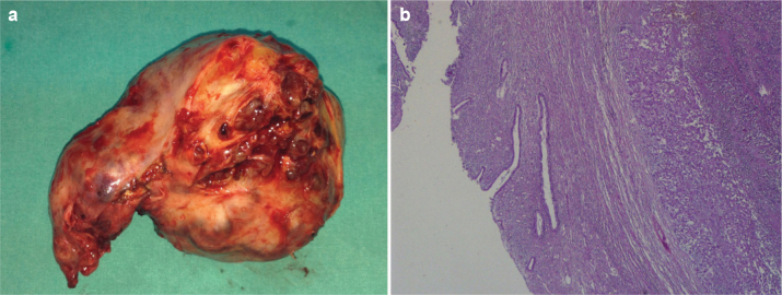
Case report: A 34-year-old woman was referred to our hospital for pelvic mass. A mass (110×100 mm) localized between the sacrococcygeal region and the uterus was detected by magnetic resonance imaging. In 2004, she had been operated upon for myxopapillary ependymoma seated in the sacrococcygeal region for the first time. She underwent tumor resection eight times due to the recurrence of spinal tumor in the same region in nine years. Under the diagnosis of uterine neoplasm, we carried out radical hysterectomy, omentectomy and pelvic lymphadenectomy as the surgical procedure. The pathological findings were reported as myxopapillary ependymoma. Immunohistochemically, the myxopapillary ependymal cells showed strong positivity for glial fibrillary acidic protein, whereas they were negative for low molecular weight cytokeratin. The Ki-67 labeling index was about 2-3%. The patient had an uneventful postoperative period. She has remained free of symptoms in the year since surgery.
Conclusion: Extra-spinal myxopapillary ependymoma is very rare, but it must be considered in the differential diagnosis of pelvic mass lesions.
Keywords: Ependymoma; spinal cord tumor; uterine cervix.
Figures


Similar articles
-
Metastases of spinal myxopapillary ependymoma: unique characteristics and clinical management.J Neurosurg Spine. 2018 Feb;28(2):201-208. doi: 10.3171/2017.5.SPINE161164. Epub 2017 Dec 8. J Neurosurg Spine. 2018. PMID: 29219779
-
Concomitant localization of a myxopapillary ependymoma at the middle thoracic part of the spinal cord and at the distal part of the filum terminale. Case report.J Neurosurg Sci. 2008 Sep;52(3):87-91. J Neurosurg Sci. 2008. PMID: 18636054
-
Remarkable efficacy of temozolomide for relapsed spinal myxopapillary ependymoma with multiple recurrence and cerebrospinal dissemination: a case report and literature review.Eur Spine J. 2018 Jul;27(Suppl 3):421-425. doi: 10.1007/s00586-017-5413-z. Epub 2017 Dec 21. Eur Spine J. 2018. PMID: 29270703 Review.
-
Surgical management of a rare myxopapillary ependymoma of the gluteal region: A case report.Surg Neurol Int. 2021 Mar 30;12:130. doi: 10.25259/SNI_768_2020. eCollection 2021. Surg Neurol Int. 2021. PMID: 33880235 Free PMC article.
-
Natural Course of Myxopapillary Ependymoma: Unusual Case Report and Review of Literature.World Neurosurg. 2019 Jan;121:239-242. doi: 10.1016/j.wneu.2018.10.028. Epub 2018 Oct 12. World Neurosurg. 2019. PMID: 30321682 Review.
Cited by
-
Myxopapillary Ependymoma Metastasis Mimicking Pulmonary Embolism: An Illustrative Case.Asian J Neurosurg. 2024 May 27;19(3):551-555. doi: 10.1055/s-0044-1779293. eCollection 2024 Sep. Asian J Neurosurg. 2024. PMID: 39205906 Free PMC article.
-
Extra-neural metastases of recurrent myxopapillary ependymoma: A patient case and literature review.Surg Neurol Int. 2025 May 16;16:182. doi: 10.25259/SNI_190_2025. eCollection 2025. Surg Neurol Int. 2025. PMID: 40469371 Free PMC article.
-
Treatment of Extraneural Metastases of Myxopapillary Ependymomas With Dose-Dense Temozolomide and Lapatinib.Cureus. 2024 Aug 27;16(8):e67928. doi: 10.7759/cureus.67928. eCollection 2024 Aug. Cureus. 2024. PMID: 39193057 Free PMC article.
References
-
- Kaner T, Sasani M, Oktenoglu T, Solmaz B, Sarloglu AC, Ozer AF. Clinical analysis of 21 cases of spinal cord ependymoma: positive clinical results of gross total resection. J Korean Neurosurg Soc. 2010;47:102–6. http://dx.doi.org/10.3340/jkns.2010.47.2.102. - DOI - PMC - PubMed
-
- Wang H, Zhang S, Rehman SK, Zhang Z, Li W, Makki MS, et al. Clinicopathological features of myxopapillary ependymoma. J Clin Neurosci. 2014;21:569–73. http://dx.doi.org/10.1016/j.jocn.2013.05.028. - DOI - PubMed
-
- Duggan MA, Hugh J, Nation JG, Robertson DI, Stuart GC. Ependymoma of the uterosacral ligament. Cancer. 1989;64:2565–71. http://dx.doi.org/10.1002/1097-0142(19891215)64:12<2565::AID-CNCR2820641.... - DOI - PubMed
-
- Mavroudis C, Townsend JJ, Wilson CB. A metastasizing ependymoma of the cauda equina. Case report. J Neurosurg. 1977;47:771–5. http://dx.doi.org/10.3171/jns.1977.47.5.0771. - DOI - PubMed
LinkOut - more resources
Full Text Sources
Other Literature Sources