. 2016 Jun 17:6:24.
doi: 10.4103/2156-7514.184244. eCollection 2016.

Fungal Infections of the Central Nervous System: A Pictorial Review

Affiliations

Fungal Infections of the Central Nervous System: A Pictorial Review

Jose Gavito-Higuera et al. J Clin Imaging Sci. .

Abstract

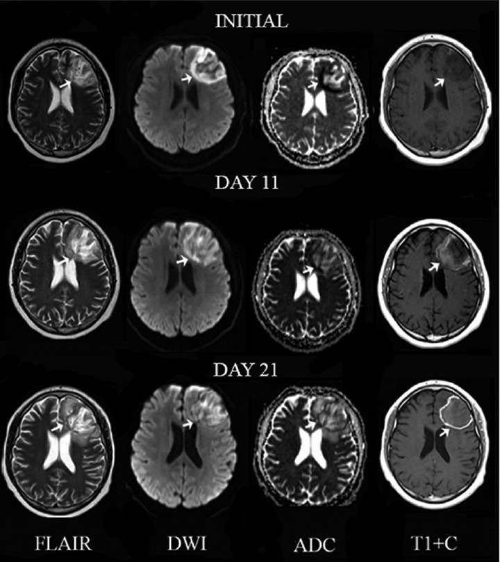
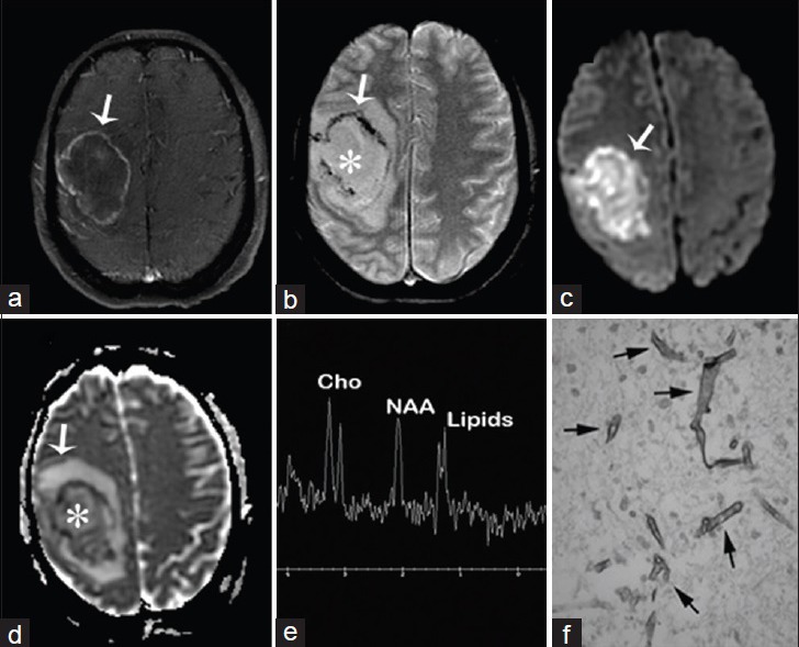
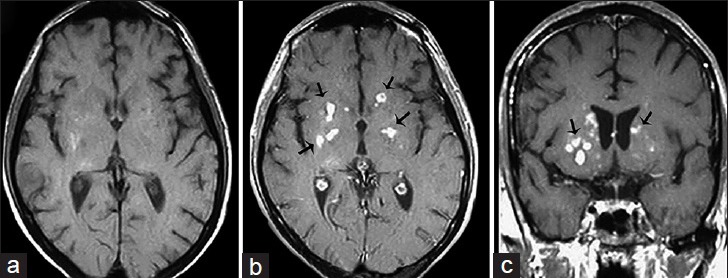
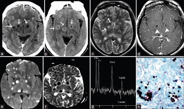
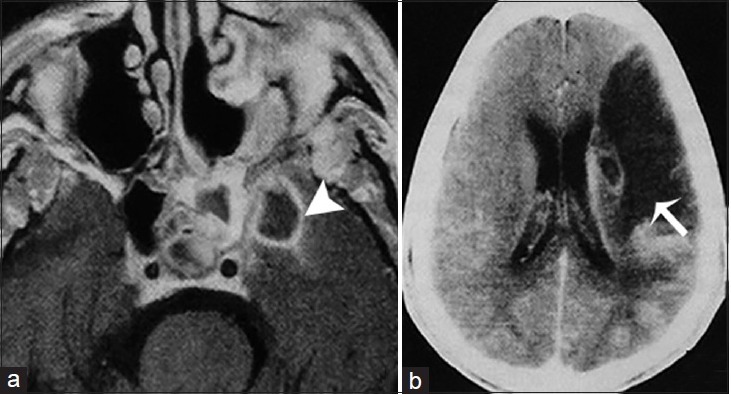
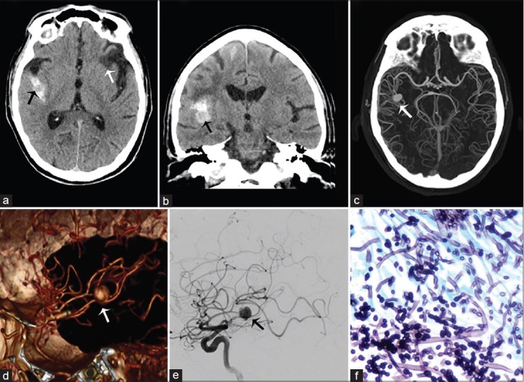
Fungal infections of the central nervous system (CNS) pose a threat to especially immunocompromised patients and their development is primarily determined by the immune status of the host. With an increasing number of organ transplants, chemotherapy, and human immunodeficiency virus infections, the number of immunocompromised patients as susceptible hosts is growing and fungal infections of the CNS are more frequently encountered. They may result in meningitis, cerebritis, abscess formation, cryptococcoma, and meningeal vasculitis with rapid disease progression and often overlapping symptoms. Although radiological characteristics are often nonspecific, unique imaging patterns can be identified through computer tomography as a first imaging modality and further refined by magnetic resonance imaging. A rapid diagnosis and the institution of the appropriate therapy are crucial in helping prevent an often fatal outcome.

Keywords: Brain abscess; cryptococcoma; fungal cerebritis; fungal meningitis; human immune deficiency virus; vascular cerebral fungal infections.


Figures

None
Jose Gavito-Higuera
Figure 1
Figure 1
A 22-year-old immunocompetent man, who presented with headaches, is diagnosed with coccidioidal meningitis. Magnetic resonance imaging T1-weighted postcontrast imaging of the brain in axial (a), coronal (b) and sagittal (c) view shows marked supra- and infra-tentorial smooth linear leptomeningeal enhancement (white arrows) is consistent with meningitis.
Figure 2
Figure 2
A 40-year-old immunocompetent man, who presented with headaches and had a history of disseminated coccidioidomycosis, is diagnosed with granulomatous leptomeningitis. Magnetic resonance imaging T1-weighted postcontrast imaging of the brain in axial (a) and sagittal (b) view shows nodular, irregular and intense leptomeningeal enhancement (arrows) involving the sylvian fissure. These findings are consistent with granulomatous leptomeningitis.
Figure 3
Figure 3
A 25-year-old man, who presented with a history of acute daily headaches over the past 6 months and had a history of chronic coccidioidal meningitis (treated), is diagnosed with chronic hypertrophic basal arachnoiditis and secondary hydrocephalus. Magnetic resonance imaging T1-weighted postcontrast imaging of the brain in axial (a) and coronal (b) view demonstrates marked leptomeningeal hypertrophy with diffuse enhancement involving predominantly the basal cisterns (arrowheads). (c) Magnetic resonance imaging fluid-attenuated inversion recovery postcontrast imaging of the brain in axial view shows a significant enlargement of the lateral ventricles with transependymal migration of cerebrospinal fluid (arrows). These findings are consistent with a hydrocephalus secondary to chronic hypertrophic basal arachnoiditis.
Figure 4
Figure 4
A 60-year-old woman, who presented with headaches, is diagnosed with coccidioidal basal arachnoiditis and acute communicating hydrocephalus. (a) Magnetic resonance imaging T1-weighted postcontrast imaging of the brain in axial view shows abnormal leptomeningeal thickening and enhancement along the perimesencephalic and superior cerebellar cisterns (arrows). (b) Magnetic resonance imaging T2-weighted imaging of the brain in axial view and magnetic resonance imaging fluid-attenuated inversion recovery (c and d) of the brain in axial and sagittal view demonstrate supratentorial ventriculomegaly associated with effacement of the cortical sulci and transependymal edema (arrowheads). These findings are consistent with basal arachnoiditis and acute communicating hydrocephalus.
Figure 5
Figure 5
A 17-year-old man with headaches and lymphoma diagnosed with Aspergillus fumigatus infection of the central nervous system. Serial magnetic resonance imaging of a left frontal lobe lesion (arrows) on T2, diffusion-weighted imaging, apparent diffusion coefficient map and T1 postcontrast (T1 + C) from left to right in axial view. Initially, the lesion appears ill-defined with increased signal intensity with foci of signal void on T2, restricted diffusion on diffusion-weighted imaging and apparent diffusion coefficient map and no abnormal enhancement on T1 post contrast suggesting late cerebritis. An 11-day follow-up shows decrease of signal voids on T2, restricted diffusion on diffusion-weighted imaging and faint peripheral enhancement on T1 post contrast consistent with early capsulitis. A 21-day follow-up demonstrates development of well-defined borders and linear peripheral enhancement on T1 post contrast consistent with late capsulitis.
Figure 6
Figure 6
A 50-year-old woman with acute myeloid leukemia, fever, headaches, and neurologic deficits diagnosed with brain abscess due to Rhizomucor pusillus (a) Magnetic resonance imaging T1 fast spin echo/fat sat postcontrast of the brain, axial view shows right sided mass with enhancing wall (arrow) (b) magnetic resonance imaging T2* GRE, axial view shows nonenhancing liquefied core (asterisk) and hypointense wall with susceptibility artifact (arrow) (c and d) diffusion-weighted imaging and apparent diffusion coefficient map of the brain, axial view show restricted diffusion of the wall and intracavitary projections (arrows) and no restriction inside the lesion (asterisk d) (e) MR spectroscopy of the brain shows inverted NAA/Cho ratio due to NAA depletion and increased lipids due to cell destruction (f) Gomori methenamine silver nitrate stain shows Zygomycetes (arrows).
Figure 7
Figure 7
A 38-year-old immunocompromised male presenting with headache and altered mental status diagnosed with Cryptococcosis (a) Magnetic resonance imaging T1-weighted imaging of the head in axial view, T1 postcontrast-weighted imaging of the head in axial (b) and coronal view (c) reveal multiple small enhancing nodules in the basal ganglia bilaterally within perivascular spaces consistent with cryptococcomas (arrows).
Figure 8
Figure 8
A 52-year-old woman, human immunodeficiency virus-positive with neurological deficits diagnosed with central nervous system cryptococcosis. (a) Noncontrast and (b) contrast-enhanced computed tomography scan of the brain, axial view show rim enhancing lesions in the basal ganglia (arrows). (c) Magnetic resonance imaging T2, axial view demonstrates increased signal intensity in the lesions. (Gelatinous pseudocysts) (arrows). (d) Magnetic resonance imaging T1 postcontrast shows rim enhancement (arrowheads), (e) B1000 data set and corresponding ADC map (f) show restricted diffusion (arrows). (g) MR spectroscopy shows inverted NAA/Cho ratio with lipids and lactate consistent with infection (h) GSM stain shows fungal organism (arrow).
Figure 9
Figure 9
A 51-year-old man, who presented with right-sided paresis and had a history of uncontrolled diabetes mellitus, is diagnosed with chronic invasive fungal Mucormycosis. (a) Magnetic resonance imaging T1 postcontrast imaging of the brain in axial view demonstrates an infectious process in the left sphenoidal sinus and an intracranial intraparenchymal ring enhancing extension in the medial temporal lobe indicative of an abscess (arrowhead). (b) Contrast-enhanced computer tomography image of the same patient shows and acute infarct in the vascular territory of the left middle cerebral artery secondary to intracranial vasculitis (arrow).
Figure 10
Figure 10
A 55-year-old man with history of intravenous drug abuse, headache, and sepsis due to a Candida sp. infection, diagnosed with mycotic aneurysm of the M2–M3 right middle cerebral artery. Computed tomography imaging of the brain without contrast in axial (a) and coronal (b) view demonstrate subarachnoid hemorrhage in the right lateral fissure surrounding right middle cerebral artery aneurysm (black arrow) and areas of encephalomalacia of prior ischemic events (white arrow). (c) Maximum intensity projection computed tomography angiogram, (d) three-dimensional reconstruction and (e) lateral selective angiogram show a saccular aneurysm arising from the M2–M3 right middle cerebral artery (arrows). (f) H and E stain with hyphae of Candida albicans.

References

    1. Mohan S, Jain KK, Arabi M, Shah GV. Imaging of meningitis and ventriculitis. Neuroimaging Clin N Am. 2012;22:557–83. - PubMed
    1. Mathur M, Johnson CE, Sze G. Fungal infections of the central nervous system. Neuroimaging Clin N Am. 2012;22:609–32. - PubMed
    1. Aiken AH. Central nervous system infection. Neuroimaging Clin N Am. 2010;20:557–80. - PubMed
    1. Tung GA, Rogg JM. Diffusion-weighted imaging of cerebritis. AJNR Am J Neuroradiol. 2003;24:1110–3. - PMC - PubMed
    1. Jain KK, Mittal SK, Kumar S, Gupta RK. Imaging features of central nervous system fungal infections. Neurol India. 2007;55:241–50. - PubMed