. 2016 Oct;76(14):1312-25.
doi: 10.1002/pros.23221. Epub 2016 Jul 12.

Neuronal Trans-Differentiation in Prostate Cancer Cells

Affiliations

Neuronal Trans-Differentiation in Prostate Cancer Cells

Andrew Farach et al. Prostate. 2016 Oct.

Abstract

Background: Neuroendocrine (NE) differentiation in prostate cancer (PCa) is an aggressive phenotype associated with therapy resistance. The complete phenotype of these cells is poorly understood. Clinical classification is based predominantly on the expression of standard NE markers.

Methods: We analyzed the phenotype of NE carcinoma of the prostate utilizing in vitro methods, in silico, and immunohistochemical analyses of human disease.

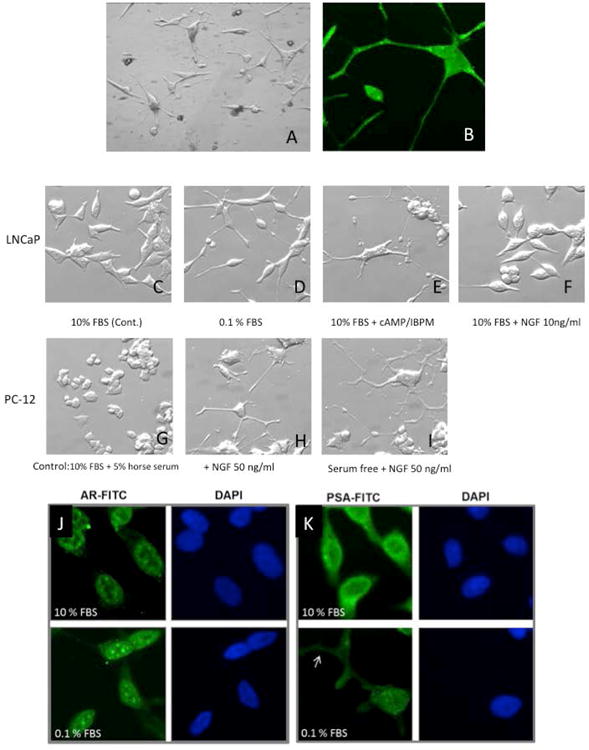
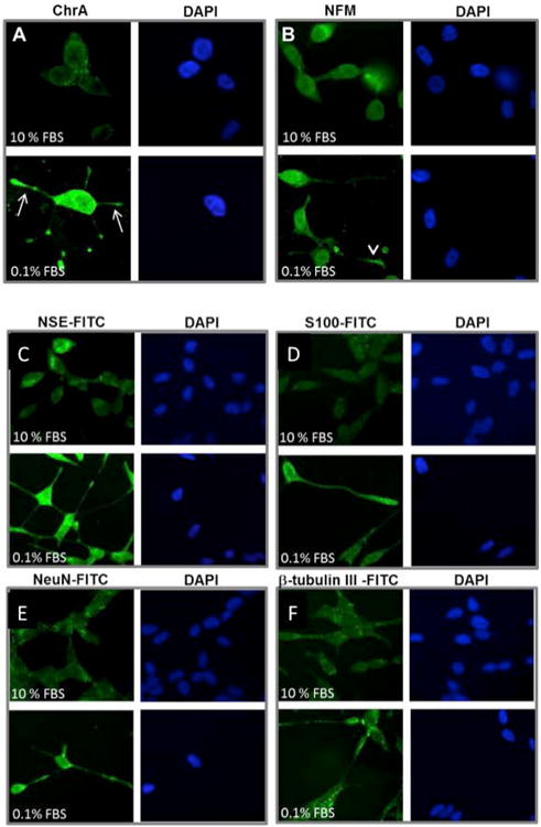
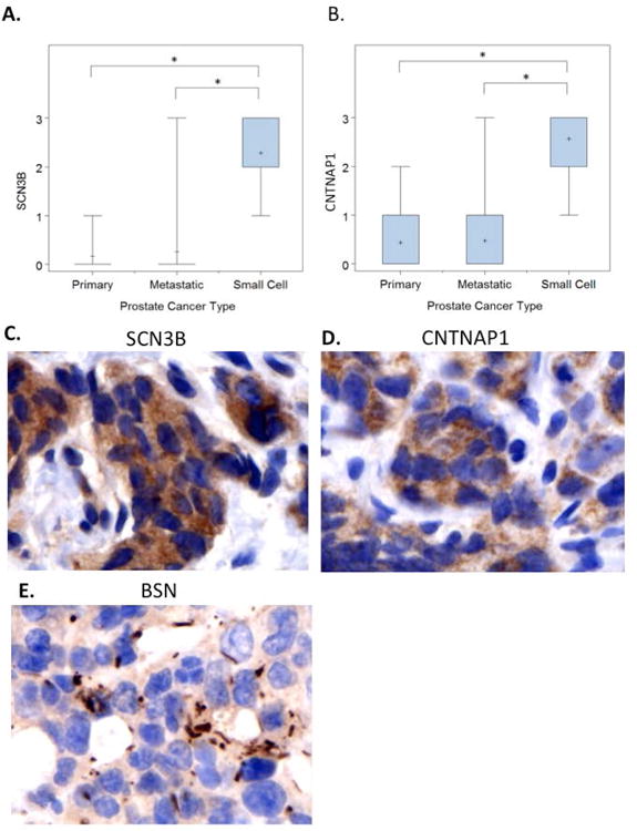
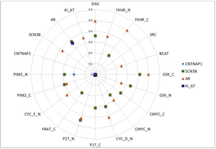
Results: LNCaP cells, subjected to a variety of stressors (0.1% [v/v] fetal bovine serum, cyclic AMP) induced a reproducible phenotype consistent with neuronal trans-differentiation. Cells developed long cytoplasmic processes resembling neurons. As expected, serum deprived cells had decreased expression in androgen receptor and prostate specific antigen. A significant increase in neuronal markers also was observed. Gene array analysis demonstrated that LNCaP cells subjected to low serum or cAMP showed statistically significant manifestation of a human brain gene expression signature. In an in silico experiment using human data, we identified that only hormone resistant metastatic prostate cancer showed enrichment of the "brain profile." Gene ontology analysis demonstrated categories involved in neuronal differentiation. Three neuronal markers were validated in a large human tissue cohort.

Conclusion: This study proposes that the later stages of PCa evolution involves neuronal trans-differentiation, which would enable PCa cells to acquire independence from the neural axis, critical in primary tumors. Prostate 76:1312-1325, 2016. © 2016 Wiley Periodicals, Inc.

Keywords: nerves; neuroendocrine; neuronal trans-differentiation; neurons; prostate cancer; resistance phenotype; small cell.


Conflict of interest statement

Conflict of Interest: The Authors have no conflicts to report that they believe could be construed as resulting in an actual, potential, or perceived conflict of interest with regard to the manuscript submitted for review.

Figures

Figure 1
Figure 1
[A] Morphological changes resembling neurons in LNCaP cells in vitro. LNCaP cells develop primary, secondary and tertiary branching, better seen in [B] immunostained with S-100. [C] LNCaP cells (Control day 5). [D] Cells demonstrate morphologic changes consistent with neuronal phenotype when stressed with low (0.1%) serum media or [E] Similar changes seen with cAMP [F]. These morphological changes were reversed in LNCaP cells with reintroduction of 10% serum. These morphological features are similar to those seen in PC12 cells [Controls in G] treated with nerve growth factor (NGF), an accepted model for neurons. [Seen in H and accentuated in I]. Immunohistochemistry to assess phenotypic changes of LNCaP cells under varying culture conditions. LNCaP cells (day 9) demonstrate [J] decreased expression of extranuclear AR and [K] decreased overall expression of PSA when cultured in serum deprived media, including in cellular processes (arrow), indicating dedifferentiation. DAPI nuclear staining is shown in blue. All images are 400x.
Figure 2
Figure 2
LNCaP cells treated with serum starvation (day 5) express higher levels of neuroendocrine markers Chromogranin A (ChrA) [A] and Neurofilament M (NF-M) [B] compared to controls in the upper level. Marker in green (FITC) and DAPI nuclear staining is shown in blue. LNCaP cells treated with serum starvation (day 5) express higher levels of neuroendocrine markers neuron specific enolase (NSE) [C] and S-100 [D]; NeuN [E] and beta tubulin [F], compared to controls in the upper level. Marker in green (FITC) and DAPI nuclear staining is shown in blue.
Figure 3
Figure 3
A: Serum starvation (0.1%FBS) and cyclic AMP treatment of LNCaP cells groups show manifestation of the human brain signature (anything above 2 could be considered significant), but not with the transfection with S4F and ASPP2. B: In tissue microarrays, 274 of 988 overexpressed genes in MET-HR are consistent with a “brain profile”, confirming the enrichment of the brain profile in metastatic hormone resistant prostate cancer. C: Brain profile obtained from publically available databases in the left panel. The right panel shows prostate epithelium and cancer of different stages from left to right: normal epithelium (EPI_NOR), epithelium adjacent to cancer (EPI_ADJ), high grade PIN (PIN), prostate cancer (PCA), hormone sensitive metastatic prostate cancer (MET_HN) and hormone resistant metastatic prostate cancer (MET_HR). Note that the enrichment of genes identified in the “brain profile” is seen only in the latter.
Figure 4
Figure 4
A and B: Semiquantitative evaluation of immunohistochemical stains for both SCN3B and CNTNAP1 show increased levels of expression in SCC of the prostate, than primary prostate cancers or non-treated metastasis. C and D: Microgranular cytoplasmic stain with antibodies against SCN3B and CNTNAP1, in brown (DAB: brown and hematoxylin background, 600×). E: Membranous/cytoplasmic structures identified with antibodies against the presynaptic protein BSN (DAB: brown and hematoxylin background, 600×).
Figure 5
Figure 5
Spearmen correlation coefficient analysis shows immunohistochemical co-localization of SCN3B and AR in tested MET-HN human tissue samples. A correlation between CNTNAP1 and Pim-2 was also noted.

Similar articles

Cited by

References

    1. Immediate versus deferred treatment for advanced prostatic cancer: initial results of the Medical Research Council Trial. The Medical Research Council Prostate Cancer Working Party Investigators Group. Br J Urol. 1997;79(2):235–46. Epub 1997/02/01. - PubMed
    1. Sharifi N, Gulley JL, Dahut WL. Androgen deprivation therapy for prostate cancer. JAMA. 2005;294(2):238–44. doi: 10.1001/jama.294.2.238. Epub 2005/07/15. - DOI - PubMed
    1. Bolla M, Collette L, Blank L, Warde P, Dubois JB, Mirimanoff RO, Storme G, Bernier J, Kuten A, Sternberg C, Mattelaer J, Lopez Torecilla J, Pfeffer JR, Lino Cutajar C, Zurlo A, Pierart M. Long-term results with immediate androgen suppression and external irradiation in patients with locally advanced prostate cancer (an EORTC study): a phase III randomised trial. Lancet. 2002;360(9327):103–6. Epub 2002/07/20. - PubMed
    1. Bolla M, Gonzalez D, Warde P, Dubois JB, Mirimanoff RO, Storme G, Bernier J, Kuten A, Sternberg C, Gil T, Collette L, Pierart M. Improved survival in patients with locally advanced prostate cancer treated with radiotherapy and goserelin. N Engl J Med. 1997;337(5):295–300. doi: 10.1056/nejm199707313370502. Epub 1997/07/31. - DOI - PubMed
    1. D'Amico AV, Manola J, Loffredo M, Renshaw AA, DellaCroce A, Kantoff PW. 6-month androgen suppression plus radiation therapy vs radiation therapy alone for patients with clinically localized prostate cancer: a randomized controlled trial. JAMA. 2004;292(7):821–7. doi: 10.1001/jama.292.7.821. Epub 2004/08/19. - DOI - PubMed

Publication types

MeSH terms