

Strong and Anomalous Thermal Expansion Precedes the Thermosalient Effect in Dynamic Molecular Crystals
- PMID: 27403616
- PMCID: PMC4941691
- DOI: 10.1038/srep29610
Strong and Anomalous Thermal Expansion Precedes the Thermosalient Effect in Dynamic Molecular Crystals
Abstract
The ability of thermosalient solids, organic analogues of inorganic martensites, to move by rapid mechanical reconfiguration or ballistic event remains visually appealing and potentially useful, yet mechanistically elusive phenomenon. Here, with a material that undergoes both thermosalient and non-thermosalient phase transitions, we demonstrate that the thermosalient effect is preceded by anomalous thermal expansion of the unit cell. The crystal explosion occurs as sudden release of the latent strain accumulated during the anisotropic, exceedingly strong expansion of the unit cell with αa = 225.9 × 10(-6) K(-1), αb = 238.8 × 10(-6) K(-1) and αc = -290.0 × 10(-6) K(-1), the latter being the largest negative thermal expansivity observed for an organic compound thus far. The results point out to the occurence of the thermosalient effect in phase transitions as means to identify new molecular materials with strong positive and/or negative thermal expansion which prior to this work could only be discovered serendipitously.
Figures


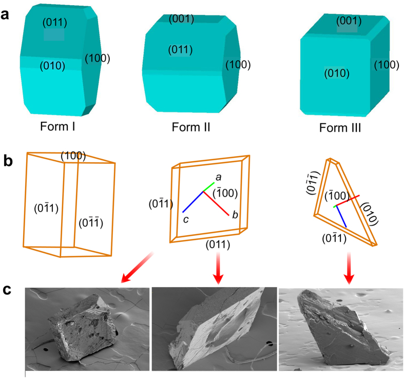

 1) face. (b) SEM images of a typical crystal glued with its (0
 1) face before (far left panel) and after the phase transition (the other panels). The circled areas are zoomed for clarity. (c) Schematic showing typical disintegration of a crystal that was glued with its (100) face onto the basis (d) SEM images before (the two panels on the far left) and after (the other panels) the transition of a typical crystal glued onto the surface with its (100) face. The dashed red arrow shows top view of the parent crystal before the transition. (e) Correlation between the mode of splitting of the crystal and the thermal expansion that causes build-up of stress.
References
-
- Wegst U. G. K., Bai H., Saiz E., Tomsia A. P. & Ritchie R. O. Bioinspired structural materials. Nature Mater. 14, 23–36 (2015). - PubMed
-
- Dicker M. P. M., Rossiter J. M., Bond I. P. & Weaver P. M. Biomimetic photo-actuation: sensing, control and actuation in sun-tracking plants. Bioinspir. Biomim. 9, 036015 (2014). - PubMed
-
- Forterre Y., Skotheim J. M., Dumais J. & Mahadevan L. How the Venus flytrap snaps. Nature 433, 421–425 (2005). - PubMed
-
- Cully A., Clune J., Tarapore D. & Mouret J.-B. Robots that can adapt like animals. Nature 521, 503–516 (2015). - PubMed
Publication types
LinkOut - more resources
Full Text Sources
Other Literature Sources
