. 2016 Jul 11:6:29492.
doi: 10.1038/srep29492.

Murine matrix metalloproteinase-20 overexpression stimulates cell invasion into the enamel layer via enhanced Wnt signaling

Affiliations

Murine matrix metalloproteinase-20 overexpression stimulates cell invasion into the enamel layer via enhanced Wnt signaling

Masashi Shin et al. Sci Rep. .

Abstract

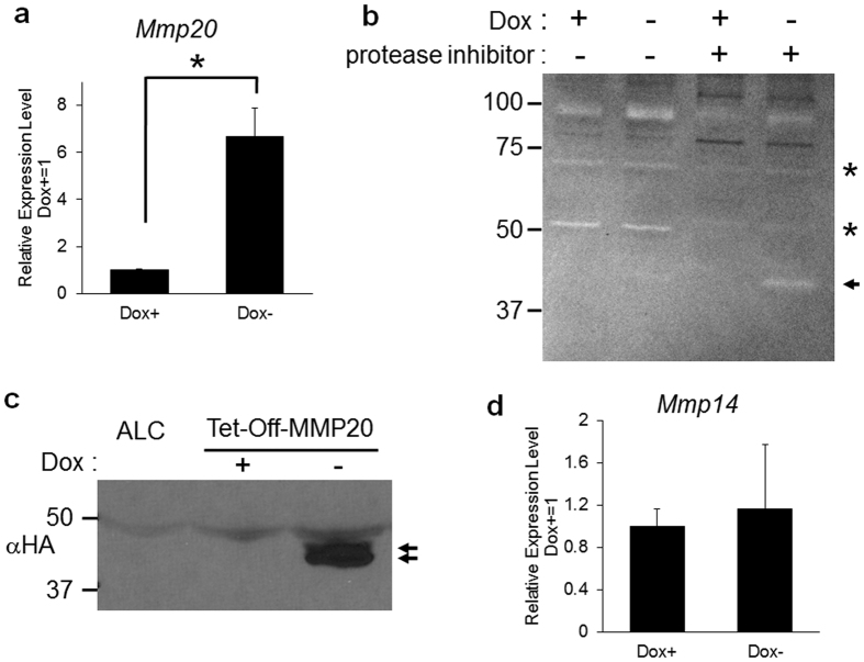
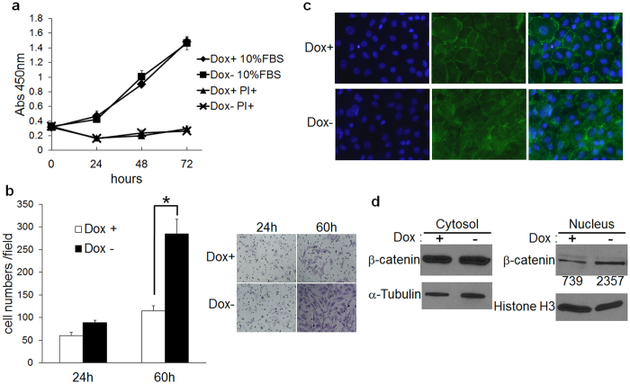
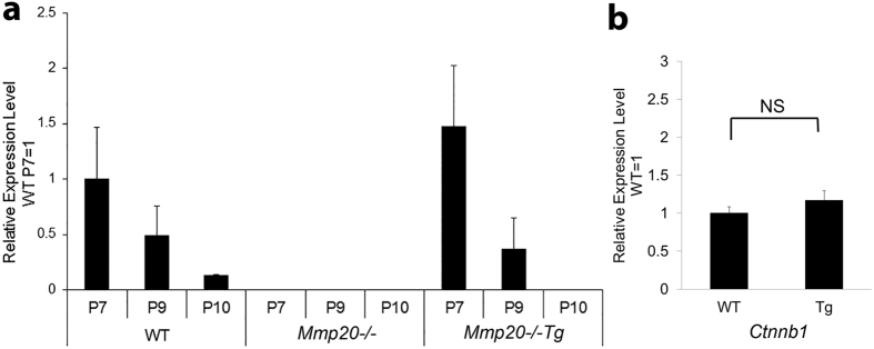
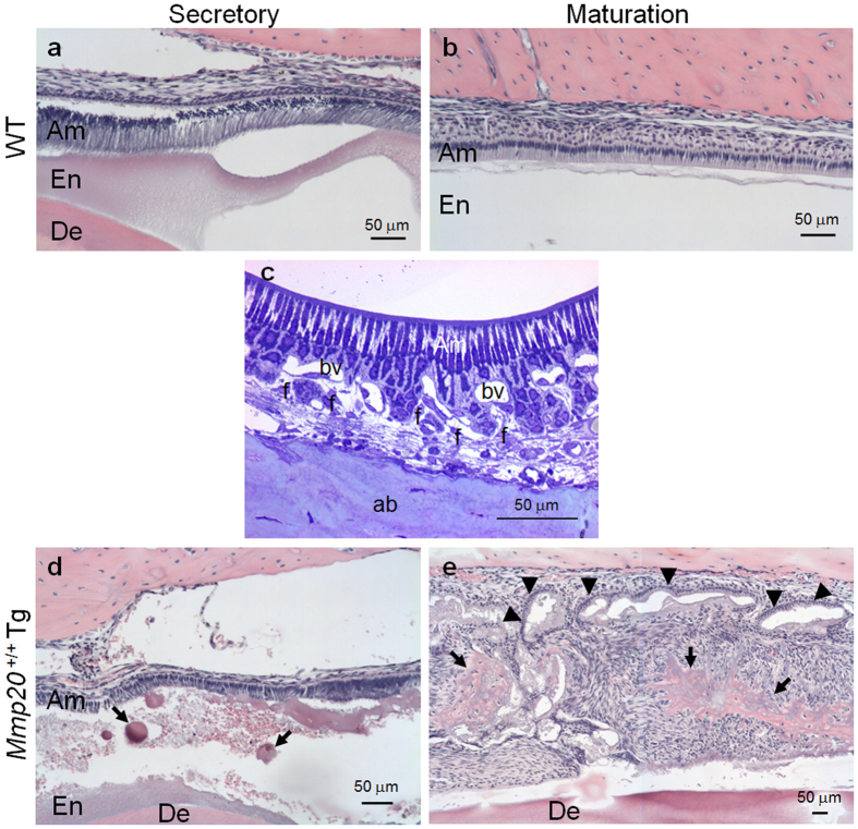
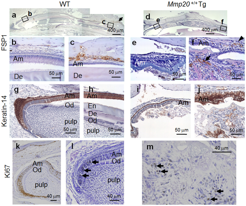
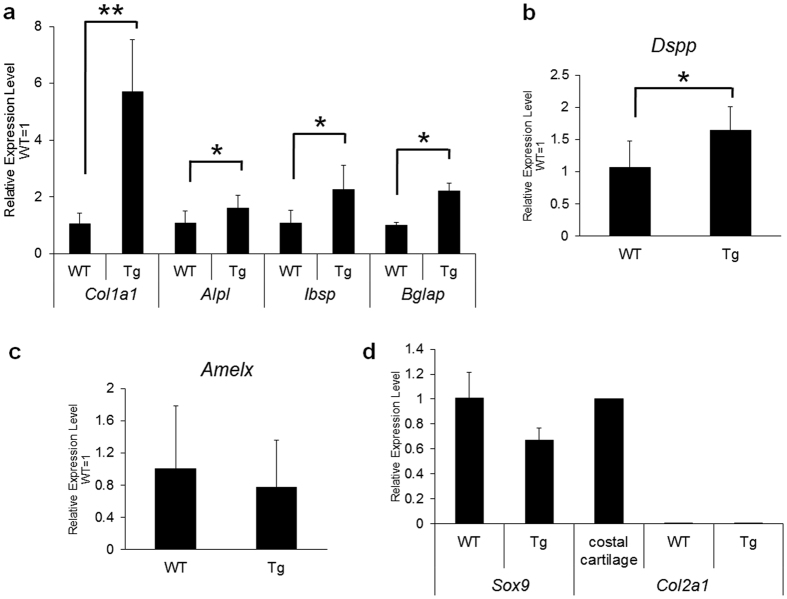
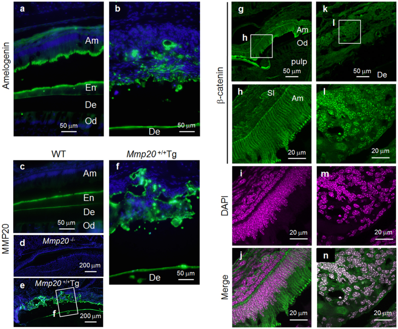
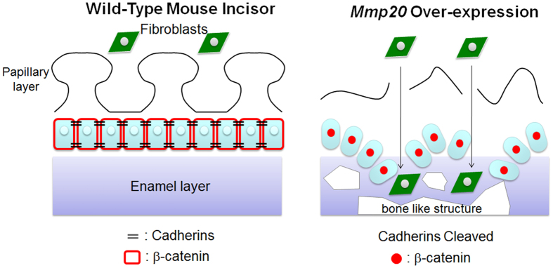
Matrix metalloproteinase-20 (MMP20) is expressed by ameloblasts in developing teeth and MMP20 mutations cause enamel malformation. We established a stably transfected Tet-Off Mmp20-inducible ameloblast-lineage cell line and found that MMP20 expression promoted cell invasion. Previously, we engineered transgenic mice (Tg) that drive Mmp20 expression and showed that Mmp20(+/+)Tg mice had soft enamel. Here we asked if Mmp20 overexpression disrupts ameloblast function. Incisors from Mmp20(+/+) mice expressing the Mmp20 Tg had a striking cell infiltrate which nearly replaced the entire enamel layer. A thin layer of enamel-like material remained over the dentin and at the outer tooth surface, but between these regions were invading fibroblasts and epithelial cells that surrounded ectopic bone-like calcifications. Mmp20(+/+)Tg mice had decreased enamel organ cadherin levels compared to the Mmp20 ablated and WT mice and, instead of predominantly locating adjacent to the ameloblast cell membrane, β-catenin was predominantly present within the nuclei of invading cells. Our data suggest that increased cadherin cleavage by transgenic MMP20 in the WT background releases excess β-catenin, which translocates to ameloblast nuclei to promote cell migration/invasion. Therefore, we conclude that MMP20 plays a role in normal ameloblast migration through tightly controlled Wnt signaling and that MMP20 overexpression disrupts this process.


Figures

Figure 1
Figure 1. An inducible MMP20-overexpressing ameloblast-lineage (ALC) cell line.
The murine Mmp20 cDNA containing a Val101-Gly101 mutation for MMP20 auto-activation and encoding a downstream hemaggultinin tag (HA) for enhanced immunodetection, was ligated into the Tet-Off Advanced Inducible gene expression vector. ALC cells were stably transfected with this vector (Tet-Off-MMP20 cells) and were assessed for levels of MMP20 expression. (a) Tet-Off-MMP20 cells were incubated with or without 100 ng/ml Doxcycline (Dox) for 48 hours followed by collection of total RNA for qPCR expression analysis. Mmp20 expression was approximately six fold higher in the Dox− as compared to the Dox+ medium (*P < 0.05). (b,c) Tet-Off-MMP20 cells were cultured in serum free medium with protease inhibitors, but not MMP inhibitors, to prevent MMP20 degradation. (b) Zymography demonstrated that in the absence of Dox, active MMP20 (arrow) was secreted into the culture medium and that protease inhibitors were necessary to visualize MMP20 activity. Asterisks denote inhibited proteases. (c) Immunoblot of the MMP20 HA tag demonstrating high level MMP20 expression (arrows) in the absence of Dox. (d) Negative control qPCR demonstrating no significant difference in Mmp14 expression levels in the presence or absence of Dox.
Figure 2
Figure 2. Analysis of cell proliferation and invasion with and without MMP20 expression.
(a) Cell proliferation was quantified by the WST-1 assay that measures dye formation by mitochondrial enzymes. No difference in cell proliferation was observed as a function of MMP20 expression over time in the presence or absence of fetal bovine serum (FBS). PI, protease inhibitors excluding MMP inhibitors. (b) Tet-Off-MMP20 cells were added to filter well inserts (8 μm pore size) coated with Matrigel and were placed into culture wells. After incubation for 24 h or 60 h in serum free medium with non-MMP protease inhibitors, the filters were washed, fixed and hematoxylin stained. Five random microscope cell fields per insert were counted under a microscope and the cell counts showed that more cells migrated to the opposite side of the filter when MMP20 was expressed (Dox−) compared to when MMP20 was not expressed (Dox+). Data are the mean ± SD for three independent experiments. *P < 0.05. The panel on the right shows example cell fields for each time and treatment. (c) Tet-Off-MMP20 cells were seeded on glass coverslips with or without 100 ng/ml Dox for 48 h. Immunofluoresence identified β-catenin location and DAPI staining identified cell nuclei. β-catenin localization adjacent to cell membranes was disrupted with MMP20 expression (Dox−). (d) Immunoblots were performed on β-catenin extracted from the cell cytosol or nucleus after 24 h treatment with or without Dox. More β-catenin was present within the cell nuclei when MMP20 was expressed (Dox−). Loading control proteins were α-tubulin for the cytosol and histone H3 for the nucleus. Numbers indicate relative band intensities after scanning.
Figure 3
Figure 3. Relative expression levels of Mmp20 in wild-type (WT) and Mmp20−/−Tg enamel organ.
Total RNA was isolated from first molar enamel organs and pulp of 7-day-old (P7, late secretory-early maturation stage), 9-day-old (P9, early-mid maturation stage) and 10-day-old (P10, mid-maturation stage) pups. (a) The mouse genotypes examined were WT, Mmp20−/− and Mmp20−/−Tg. Mmp20 expression was quantified by qPCR and as expected, no expression was observed in the Mmp20−/− mice. In WT mice, Mmp20 was expressed in the enamel organ and expression declined with each successive developmental stage. The amelogenin promoter driven transgene in the Mmp20−/−Tg had an Mmp20 expression pattern similar to that of WT. (b) β-catenin (Ctnnb1) expression levels were not significantly (NS) increased in enamel organs from Mmp20+/+Tg (Tg) mice compared to WT.
Figure 4
Figure 4. Identification of a striking cell infiltrate present within the Mmp20+/+Tg mouse incisor enamel space.
Demineralized longitudinal incisor sections from (a) wild-type (WT) secretory and (b) WT maturation stages. (c) Semithin sections of plastic embedded samples consisting of maturation stage enamel organs from WT mice were stained with toluidine blue. The outer surface of the papillary layer of the enamel organ is intimately associated with blood vessels (bv) and other connective tissue elements including fibroblasts (f) extending out to the labial alveolar bone (ab). (d) In contrast to WT, the Mmp20+/+Tg secretory stage incisors had ectopic calcifications in the enamel space (arrows). (e) The Mmp20+/+Tg maturation stage incisors had a profound pathology consisting of a massive cell infiltrate, extensive ectopic calcifications (arrows) and a disorganized ameloblast layer that appeared to weave in and out of the sectioning plain (arrow heads).
Figure 5
Figure 5. Localization of fibroblast specific protein-1 (FSP1) and Keratin-14 (K14) positive cells, and identification of proliferating cells via Ki67 expression in wild-type (WT) and Mmp20+/+Tg mouse incisors.
Immunostaining for FSP1 was performed on longitudinal incisor sections. (ac) Incisor sections from WT mice. FSP1 positive cells were detected in tissues near the (a, arrow) lips and in the loose connective tissues and (b,c) vacular spaces near labial bone. (df) Incisor sections from Mmp20+/+Tg mice. (d) The cell infiltrate in the Tg incisors became abundant soon after what appears to be the mid-late secretory stage (e,f). Within the cell infiltrate, FSP1 positive cells were present near the (f, arrow) ectopic bone-like mineral and near the (f, arrowhead) labial alveolar bone. Immunohistochemical staining of K14 was performed on longitudinal incisor sections from (g,h) WT and (i,j) Mmp20+/+Tg mice. (g) In WT mice, the enamel organ, but not the pulp stained positively for K14. Closer inspection revealed that pre-ameloblasts did not express K14. However, (h) maturation stage ameloblasts were K14 positive. (i,j) In Mmp20+/+Tg mice, K14 was present in the enamel organ, but was not strong until the equivalent, by tooth length, of what is the maturation stage of development in WT mice. (i) The secretory stage stratum intermedium stained strongly and the ameloblasts stained lightly. (j) However, during the maturation stage, the entire enamel organ was strongly positive for K14 and the cell infiltrate also had islands of strong staining. (k) In WT 3-day-old second molars and (l) WT adult incisors, Ki67 expression was observed in nuclei of pre-ameloblasts at the apical cervical loop (arrows). In contrast, (m) in Mmp20+/+Tg incisors, the cell infiltrate contained cells positive for nuclear Ki67 expression (arrows) indicating that a portion of the infiltrated cells were undergoing cell division. Ameloblasts, Am; odontoblasts, Od; enamel space, En and dentin, De.
Figure 6
Figure 6. Expression levels of bone-related genes in wild-type (WT) and Mmp20+/+Tg (Tg) mouse incisor enamel.
Whole incisors were ground, extracted for total RNA and prepared for qPCR quantification of gene expression. (a) Significantly increased transcript levels of alkaline phosphatase (Alpl), osteocalcin (Bglap), bone sialoprotein (Ibsp) and a highly significant increase in collagen 1α1 chain (Col1a1) transcripts were observed in incisors from Mmp20+/+Tg mice versus WT mice. (bd) A significant increase in expression was observed for dentin sialophosphoprotein (Dspp), but not for amelogenin (Amelx), Sox9 or collagen 2α1 (Col2a1) in the Tg mice relative to the WT controls. These expression differences were consistent with formation of bone-like calcifications observed within the enamel space of the Tg mice. *P < 0.05; **P < 0.01.
Figure 7
Figure 7. Amelogenin and MMP20 expression plus β-catenin localization in wild-type (WT) and Mmp20+/+Tg incisors.
(a) Immnofluorescence localization confirmed that WT secretory stage ameloblasts express amelogenin and demonstrated that (b) cell islands within the cell infiltrate of Mmp20+/+Tg incisors also express amelogenin. (c) WT, but not (d) Mmp20−/− secretory stage ameloblasts express MMP20. Interestingly, in Mmp20+/+Tg incisors, the MMP20 expression pattern (e,f) was similar to the amelogenin expression pattern (b) indicating that the cell infiltrate was composed of cells that may formerly have been secretory stage ameloblasts or their derivatives. Immunofluorescence was performed with antibodies specific for β-catenin and DAPI staining (color was changed from blue to magenta) identified the nuclei in WT and Mmp20+/+Tg incisors. (gj) β-catenin located along the cell membrane in WT ameloblasts, while (kn) β-catenin predominantly located to nuclei within Mmp20+/+Tg ameloblasts. These data suggest that in Mmp20+/+Tg incisors, cadherin cleavage by MMP20 releases β-catenin from its predominant position adjacent to the plasma membrane and that β-catenin relocates to cell nuclei to promote cell invasion into the enamel space. Ameloblasts, Am; stratum intermedium, SI; enamel space, En; dentin, De; odontoblasts, Od.
Figure 8
Figure 8. Schematic depicting how fibroblasts and epithelial cells may gain entry into the Mmp20+/+Tg incisor enamel space.
Mmp20 overexpression results in increased cadherin cleavage, which may disrupt the ameloblast layer. When the ameloblast layer is disrupted, fibroblasts near the capillary basement membranes within the papillary layer may become free to move into the enamel space. These invading fibroblasts may contribute to the formation of the ectopic bone-like formations found within the Mmp20+/+Tg incisor enamel space. Epithelial and fibroblast cell migration into the enamel space may be enhanced by β-catenin translocation to cell nuclei due to extensive cadherin cleavage and resultant β-catenin release by the MMP20 overexpressing mice.

Similar articles

Cited by

References

    1. Roycik M. D., Fang X. & Sang Q. X. A fresh prospect of extracellular matrix hydrolytic enzymes and their substrates. Curr Pharm Des 15, 1295–1308 (2009). - PubMed
    1. Bartlett J. D., Ryu O. H., Xue J., Simmer J. P. & Margolis H. C. Enamelysin mRNA displays a developmentally defined pattern of expression and encodes a protein which degrades amelogenin. Connect Tissue Res 39, 101–109, discussion 141-109 (1998). - PubMed
    1. Bartlett J. D. & Simmer J. P. Proteinases in developing dental enamel. Crit Rev Oral Biol Med 10, 425–441 (1999). - PubMed
    1. Fukae M. et al.. Enamelysin (matrix metalloproteinase-20): localization in the developing tooth and effects of pH and calcium on amelogenin hydrolysis. J Dent Res 77, 1580–1588 (1998). - PubMed
    1. Begue-Kirn C., Krebsbach P. H., Bartlett J. D. & Butler W. T. Dentin sialoprotein, dentin phosphoprotein, enamelysin and ameloblastin: tooth-specific molecules that are distinctively expressed during murine dental differentiation. Eur J Oral Sci 106, 963–970 (1998). - PubMed

Publication types

LinkOut - more resources