. 2016 Jul 1;57(8):3665-73.
doi: 10.1167/iovs.16-19114.

Nox4 Plays a Role in TGF-β-Dependent Lens Epithelial to Mesenchymal Transition

Affiliations

Nox4 Plays a Role in TGF-β-Dependent Lens Epithelial to Mesenchymal Transition

Shannon J Das et al. Invest Ophthalmol Vis Sci. .

Abstract

Purpose: Transforming growth factor-β induces an epithelial to mesenchymal transition (EMT) in the lens, presented as an aberrant growth and differentiation of lens epithelial cells. Studies in other models of EMT have shown that TGF-β-driven EMT is dependent on the expression of the reactive oxygen species (ROS)-producing enzyme nicotinamide adenine dinucleotide phosphate (NADPH)-oxidase-4 (Nox4). We investigate the role of this enzyme in TGF-β-induced lens EMT and determine whether it is required for this pathologic process.

Methods: Rat lens epithelial explants were used to investigate the role of Nox4 in TGF-β-driven lens EMT. Nox1-4 expression and localization was determined by immunolabeling and/or RT-PCR. NADPH-oxidase-produced ROS were visualized microscopically using the fluorescent probe, dihydroethidium (DHE). VAS2870, a pan-NADPH oxidase inhibitor, was used to determine the specificity of Nox4 expression and its role in ROS production, and subsequently TGF-β-driven EMT.

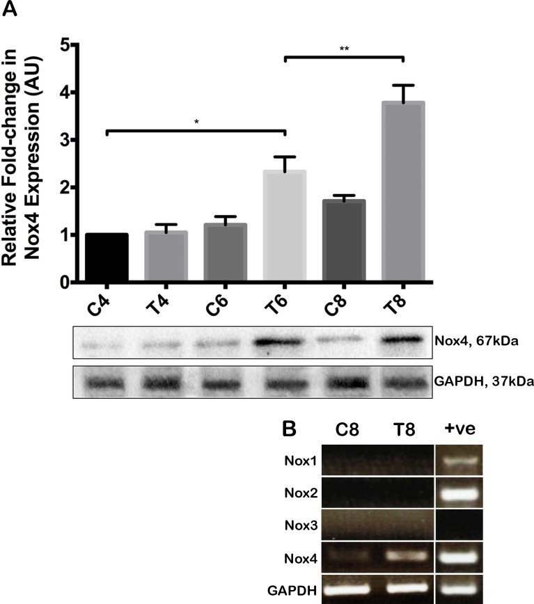
Results: We demonstrate, for the first time to our knowledge, in rat lens epithelial explants that TGF-β treatment induces Nox4 (but not Nox1-3) expression and activity. Increased Nox4 expression was first detected at 6 to 8 hours following TGF-β treatment and was maintained in explants up to 48 hours. At 8 hours after TGF-β treatment, Nox4 was observed in cell nuclei, while at later stages in the EMT process (at 48 hours), Nox4 was predominately colocalized with α-smooth muscle actin. The inhibition of Nox4 expression and activity using VAS2870 inhibited EMT progression.

Conclusions: Transforming growth factor-β drives the expression of the ROS-producing enzyme Nox4 in rat lens epithelial cells and Nox4 inhibition can impede the EMT process.


Figures

Figure 1
Figure 1
Transforming growth factor-β2 induces an increase in Nox4 expression in lens epithelial explants: Western blot. (A) Representative Western blot depicting the time-course (4, 6, and 8 hours) of Nox4 expression following TGF-β2 treatment (T4, T6, and T8), compared to control explants (C4, C6, and C8). Image is representative of 3 independent experiments. Above: Densitometry was performed to normalize the expression of Nox4 protein to GAPDH expression. Upon TGF-β2 treatment, Nox4 expression was significantly upregulated at 6 hours (T6), and these levels significantly increased by 8 hours (T8). Data are presented as mean ± SEM. One-way ANOVA, Tukey's post hoc test (2-tailed t-test) *P < 0.05 and **P < 0.01. (B) A representative RT-PCR of Nox1–4 mRNA transcripts after 8 hours, in the absence (C8) and presence (T8) of TGF-β2. Positive controls are included for Nox1, 2, and 4. For each lane, the PCR products shown correspond to the expected base pair length (Nox1, 201 base pairs [bp]; Nox2, 149 bp; Nox3, 234 bp; Nox4, 191 bp, and GAPDH, 287 bp).
Figure 2
Figure 2
Transforming growth factor-β2 induces variable Nox4 localization patterns in lens epithelial cells with the progression of TGF-β induced EMT. Nox4 (red) was merged with the cell nuclear Hoechst dye (blue). Untreated explants at 8 (A) and 48 (C) hours exhibited little Nox4 expression. Explants treated with TGF-β2 (+TGF-β2) exhibited a stronger distinct punctate label by 8 hours (B) that primarily localized to the perinuclear regions of the cells. At 48 hours (D), this label became more pronounced throughout the cytosol. By 48 hours compared to control explants (E), epithelial cells treated with TGF-β2 also labeled for α-SMA (green; [F], with α-SMA labeled-stress fibres colocalizing with Nox4 (shown at higher magnification, Inset 1). Punctate cytosolic distribution of Nox4, independent of stress fibers, also was apparent in some cells ([F], shown at higher magnification, Inset 2). Images are representative of 3 independent experiments, consisting of 3 replicates per group. Scale bar: 40 and 20 μm for the inserts.
Figure 3
Figure 3
The ROS probe, DHE, was used to assess increases in ROS following TGF-β2 treatment. In untreated explants, basal levels of ROS were observable (AD). Importantly, TGF-β2 treated explants yielded noticeable increases in ROS by 6 hours, that increased up to 8 hours (H). Treatment with VAS2870 blocked TGF-β2–induced ROS (I) as shown by diminished fluorescence intensity, with less ROS produced. This was in conjunction to VAS2870 also blocking Nox4 protein expression (see [I] inset). Representative images from 3 independent experiments, consisting of at least 2 replicates for each treatment group. Scale bar: 100 μm (AI) and 20μm for (inset [I]). Total fluorescence intensity also was calculated using ImageJ (J), with significant increases after 6 and 8 hours, effectively blocked by VAS2870. Data are presented as mean ± SEM. One-way ANOVA, Tukey's post hoc test (2-tailed t-test) ***P < 0.001 and ****P < 0.0001.
Figure 4
Figure 4
Suppression of TGF-β2-induced EMT using VAS2870. Phase contrast images of explants at days 2 (B, F), 3 (C, G), and 5 (A, E, D, H). Explants treated with TGF-β2 alone (BD) underwent morphologic changes associated with EMT compared to untreated cells (A). Explants treated with VAS2870 and TGF-β2 (FH) showed a delayed EMT response, with less cell elongation apparent by Day 2 (F), reduced cell loss by Day 3 (G) and the retention of epithelial-like cells by Day 5 (H). Note that, VAS2870 alone (E) had little effect on cells treated up to 5 days of culture. Images are representative of 3 independent experiments, consisting of 3 replicates per group. Scale bar: 100 μm. (I) Percentage cell loss was calculated using the threshold feature of ImageJ in relation to areas of bare lens capsule, with significant cell loss in TGF-β2–treated explants from day 3, which was abrogated by application of VAS2870. Data are presented as mean ± SEM. Two-way ANOVA, Tukey's post hoc test (2-tailed t-test) **P < 0.01 and ****P < 0.0001.
Figure 5
Figure 5
Maintenance of E-cadherin expression in cells cotreated with TGF-β2 and VAS2870. Explants were treated with VAS2870 (A), or cotreated with TGF-β2 and VAS2870 (B) and fixed at 5 days of culture. Immunolabeling for the membrane marker E-cadherin (green) was performed and nuclei were counterstained with Hoechst (blue). In cells treated only with VAS2870 (A), E-cadherin localized to the cell membrane and there was no apparent cell loss. Cotreatment with TGF-β2 and VAS2870 promoted increased cell survival and the maintenance of a membranous E-cadherin label (B).
Figure 6
Figure 6
VAS2870 blocks TGF-β2–induced Nox4 and α-SMA expression. Explants cultured for 24 hours in the presence of TGF-β2 (AC), or TGF-β2 and VAS2870 (DF) were immunolabeled for Nox4 ([B, E], red), α-SMA (green; [C, F]) and counterstained with Hoechst dye (A, D). Compared to explants treated with TGFβ alone (AC), VAS2870 was shown to block TGF-β2-induced Nox4 (E) and α-SMA (F) expression. Images are representative of 3 independent experiments, consisting of 3 replicates per group. Scale bar: represents 50 μm. (G) Protein lysates also were collected from explants treated with and without TGF-β2 and VAS2870 and Western blotting demonstrated similar reductions in Nox4 and α-SMA expression in the presence of VAS2870 at 24 and 48 hours. GAPDH expression remained constant regardless of treatment.

References

    1. Kubo E,, Fatma N,, Akagi Y,, Beier DR,, Singh SP,, Singh DP. TAT-mediated PRDX6 protein transduction protects against eye lens epithelial cell death and delays lens opacity. Am J Physiol Cell Physiol. 2008; 294: C842–C855. - PubMed
    1. Wu H,, Lin L,, Giblin F,, Ho YS,, Lou MF. Glutaredoxin 2 knockout increases sensitivity to oxidative stress in mouse lens epithelial cells. Free Radic Biol Med. 2011; 51: 2108–2117. - PMC - PubMed
    1. Fan X,, Liu X,, Hao S,, Wang B,, Robinson ML,, Monnier VM. The LEGSKO mouse: a mouse model of age-related nuclear cataract based on genetic suppression of lens glutathione synthesis. PLoS One. 2012; 7: e50832. - PMC - PubMed
    1. Bhuyan KC,, Bhuyan DK. Molecular cataractogenesis: III. Toxic metabolites of oxygen as initiators of lipid peroxidation and cataract. Curr Eye Res. 1984; 3: 67–81. - PubMed
    1. Kleiman NJ,, Spector A. DNA single strand breaks in human lens epithelial cells from patients with cataract. Curr Eye Res. 1993; 12: 423–431. - PubMed

Publication types

MeSH terms