Review
. 2016 Sep;110(1-2):10-18.
doi: 10.1016/j.jphysparis.2016.07.001. Epub 2016 Jul 9.

Cholinergic genetics of visual attention: Human and mouse choline transporter capacity variants influence distractibility

Affiliations
Review

Cholinergic genetics of visual attention: Human and mouse choline transporter capacity variants influence distractibility

Martin Sarter et al. J Physiol Paris. 2016 Sep.

Abstract

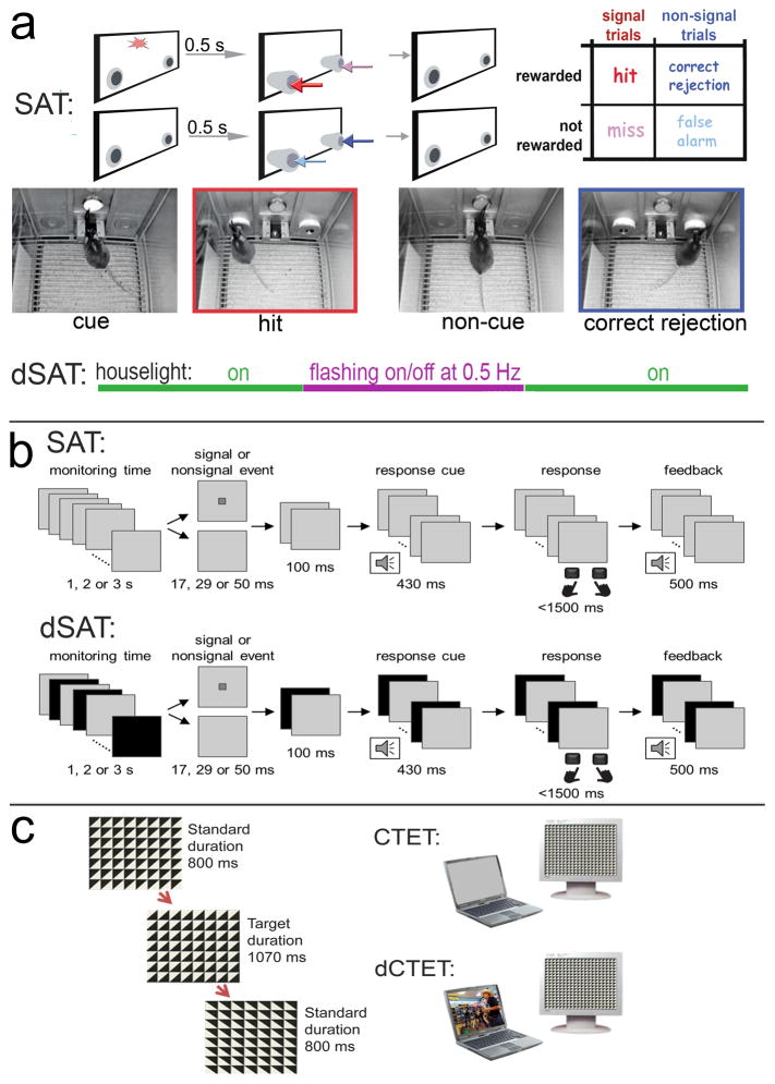
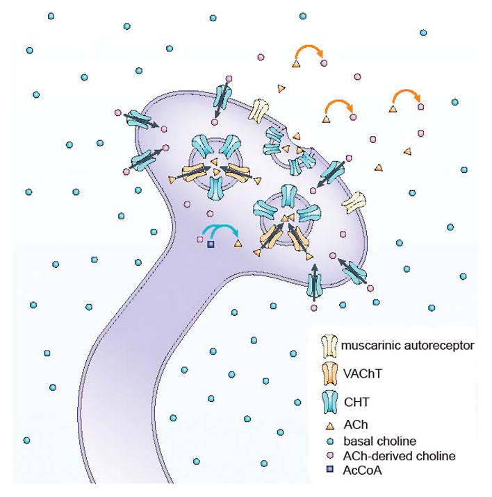
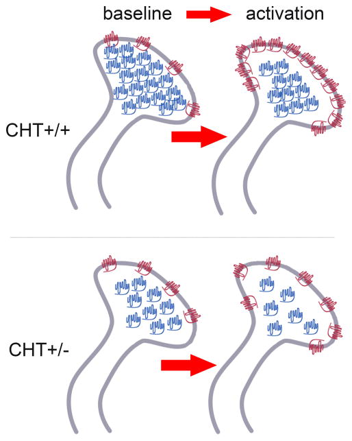
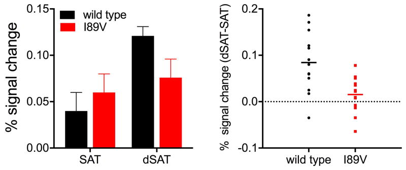
The basal forebrain cholinergic projection system to the cortex mediates essential aspects of visual attention performance, including the detection of cues and the response to performance challenges (top-down control of attention). Higher levels of top-down control are mediated via elevated levels of cholinergic neuromodulation. The neuronal choline transporter (CHT) strongly influences the synthesis and release of acetylcholine (ACh). As the capacity of the CHT to import choline into the neuron is a major, presynaptic determinant of cholinergic neuromodulation, we hypothesize that genetically-imposed CHT capacity variation impacts the balance of bottom-up versus top-down control of visual attention. Following a brief review of the cognitive concepts relevant for this hypothesis, we describe the key results from our research in mice and humans that possess genetically-imposed changes in choline uptake capacity. CHT subcapacity is associated with poor top-down attentional control and attenuated (cholinergic) activation of right frontal regions. Conversely, mice overexpressing the CHT, and humans expressing a CHT variant hypothesized to enhance choline transporter function, are relatively resistant to challenges of visual attention performance. Genetic or environmental modulation of CHT expression and function may be associated with vulnerabilities for cognitive disorders.

Keywords: Acetylcholine; Attention; Choline transporter; Genetics; Humans; Rodents.


Figures

Figure 1
Figure 1. Tasks used in rodents and humans to assess bottom-up and top-down attention
a: The rodent Sustained Attention Task (SAT) consists of a randomized sequence of cue and non-cue events, followed by extension of the two response ports (McGaughy et al., 1996; McGaughy and Sarter, 1995; St Peters et al., 2011a). Hits and correct rejections, but not misses and false alarms, are rewarded. Note the matching colors of the arrows pointing to the response ports for the four response categories and the arrows in the outcome matrix on the right, indicating that, in cued trials, hits (red arrows) are rewarded and in non-cued trials, correct rejections are rewarded (dark blue). Conversely, misses (pink arrows) and false alarms (light blue arrows) are not rewarded and trigger the intertrial interval. The photographs depict the mouse version of the task and show the onset of a cued (left) and non-cued trial (third from left), and a hit (red frame, matching arrow color) and a correct rejection (dark blue frame). dSAT: To test top-down control mechanisms, a disruptor (houselights flashing at 0.5 Hz; dSAT) impairs performance, followed by performance recovery. The disruptor is presented during a period in the middle of a regular session (see violet line), typically for 16 min of a 60 min test session. SAT performance is associated with significant increases in levels of cholinergic neuromodulation in the cortex, and dSAT performance further elevates cholinergic activity and this increase correlates positively with post-distractor recovery (St Peters et al., 2011b). b: The human SAT, similar to the rodent task, requires reporting the presence or absence of a cue, and in the dSAT the computer screen flashes continuously from gray to black at a rate of 10 Hz (Demeter et al., 2013; Demeter et al., 2008). Human subjects increase right frontal activity (BA9) while performing the dSAT, indicating their persistent reliance on top-down mechanisms (Demeter et al., 2011). c: The CTET (Continuous Temporal Expectancy Test; O’Connell et al., 2009) consists of a black and white grid made up of squares divided into triangles. The triangles rotate randomly (90°, 180°, or 270°) and consecutive rotations are separated by 800 ms. Participants press the spacebar when noting that a longer period (1070 ms) separated consecutive rotations. The distractor-CTET uses a laptop oriented 32° to the left of the main task computer. The laptop is silent and displays a gray screen during CTET, and it plays content-rich video clips with sound during dCTET.
Figure 2
Figure 2. Schematic illustration of the main steps of presynaptic cholinergic transmission
The cholinergic terminal is situated in the extracellular space, which contains low (micromolar) and relatively stable concentrations of basal choline. Hydrolysis of acetylcholine (ACh) (orange arrows) gives rise to ACh-derived choline, which is then effectively recycled into the presynaptic terminals by hemicholinium-3-sensitive, high-affinity choline uptake transporters (CHT). ACh-derived choline and acetyl-coenzyme A together give rise to ACh in the presynaptic terminal, catalysed by choline acetyltransferase (ChAT; blue arrows). The illustration is not intended to suggest that only choline molecules that are derived from ACh are transported back into the terminal; rather, it reflects evidence indicating that amounts proportional to ACh-derived choline are rapidly cleared from the extracellular space. The illustration does not depict the low-affinity transporter and the use of choline for other purposes, such as phospholipid metabolism (Lockman and Allen, 2002). Vesicular acetylcholine transporters pack newly synthesized ACh into vesicles. Because most CHTs are located on the vesicular membrane, changes in the activity of cholinergic terminals are predicted to alter the trafficking of CHTs between the plasma and vesicular membrane (Ferguson et al., 2004; Ferguson et al., 2003; Simon et al., 1976). (Figure and legend reproduced, in accordance with NPG Licence Policy, from Sarter and Parikh, 2005.)
Figure 3
Figure 3. Symbolic Illustration of the effects of BF stimulation or attentional performance on the subcellular synaptosomal distribution of CHTs
At baseline, comparable densities of synaptosomal plasma membrane CHTs (depicted in red) in CHT+/+ and CHT+/− mice support similar levels of basal ACh release and choline uptake. CHT heterozygosity primarily reduces the density of intracellular CHTs (in blue). Therefore, stimulation of cholinergic neurons or attentional performance reveal the impact of CHT heterozygosity, yielding attenuated levels of CHTs in plasma membrane and thus attenuated levels of cholinergic neuromodulation. (Adopted from Parikh et al., 2013, and reproduced in accordance with the J. Neurosci. Licence to Publish.)
Figure 4
Figure 4. I89V subjects fail to activate right PFC in response to challenge
Controls, but not I89V humans increase right PFC activation in response to attentional challenge. Percent signal change in the bar graphs (left) is reported relative to fixation baseline. Individual participant data (right) is plotted as percent signal change for the index dSAT-SAT. A significant group by disruptor interaction indicated controls increased activation in right PFC but I89V did not (for details see Berry et al., 2015).

Similar articles

Cited by

References

    1. Apparsundaram S, Martinez V, Parikh V, Kozak R, Sarter M. Increased capacity and density of choline transporters situated in synaptic membranes of the right medial prefrontal cortex of attentional task-performing rats. J Neurosci. 2005;25(15):3851–3856. - PMC - PubMed
    1. Bazalakova MH, Blakely RD. The high-affinity choline transporter: a critical protein for sustaining cholinergic signaling as revealed in studies of genetically altered mice. Handb Exp Pharmacol. 2006;(175):525–544. - PubMed
    1. Bazalakova MH, Wright J, Schneble EJ, McDonald MP, Heilman CJ, Levey AI, Blakely RD. Deficits in acetylcholine homeostasis, receptors and behaviors in choline transporter heterozygous mice. Genes Brain Behav. 2007;6(5):411–424. - PubMed
    1. Berry AS, Blakely RD, Sarter M, Lustig C. Cholinergic capacity mediates prefrontal engagement during challenges to attention: evidence from imaging genetics. Neuroimage. 2015;108:386–395. - PMC - PubMed
    1. Berry AS, Demeter E, Sabhapathy S, English BA, Blakely RD, Sarter M, Lustig C. Disposed to distraction: genetic variation in the cholinergic system influences distractibility but not time-on-task effects. J Cogn Neurosci. 2014a;26(9):1981–1991. - PMC - PubMed