

doi: 10.1021/acs.biochem.6b00626.
Epub 2016 Jul 22.
Crystal Structures of the Iron-Sulfur Cluster-Dependent Quinolinate Synthase in Complex with Dihydroxyacetone Phosphate, Iminoaspartate Analogues, and Quinolinate
Affiliations
- PMID: 27404889
- PMCID: PMC7775720
- DOI: 10.1021/acs.biochem.6b00626
Item in Clipboard
Crystal Structures of the Iron-Sulfur Cluster-Dependent Quinolinate Synthase in Complex with Dihydroxyacetone Phosphate, Iminoaspartate Analogues, and Quinolinate
Biochemistry.
.
Abstract
The quinolinate synthase of prokaryotes and photosynthetic eukaryotes, NadA, contains a [4Fe-4S] cluster with unknown function. We report crystal structures of Pyrococcus horikoshii NadA in complex with dihydroxyacetone phosphate (DHAP), iminoaspartate analogues, and quinolinate. DHAP adopts a nearly planar conformation and chelates the [4Fe-4S] cluster via its keto and hydroxyl groups. The active site architecture suggests that the cluster acts as a Lewis acid in enediolate formation, like zinc in class II aldolases. The DHAP and putative iminoaspartate structures suggest a model for a condensed intermediate. The ensemble of structures suggests a two-state system, which may be exploited in early steps.
Figures
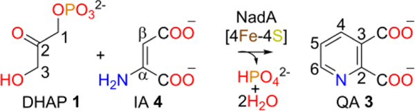
NadA catalyzed reaction.
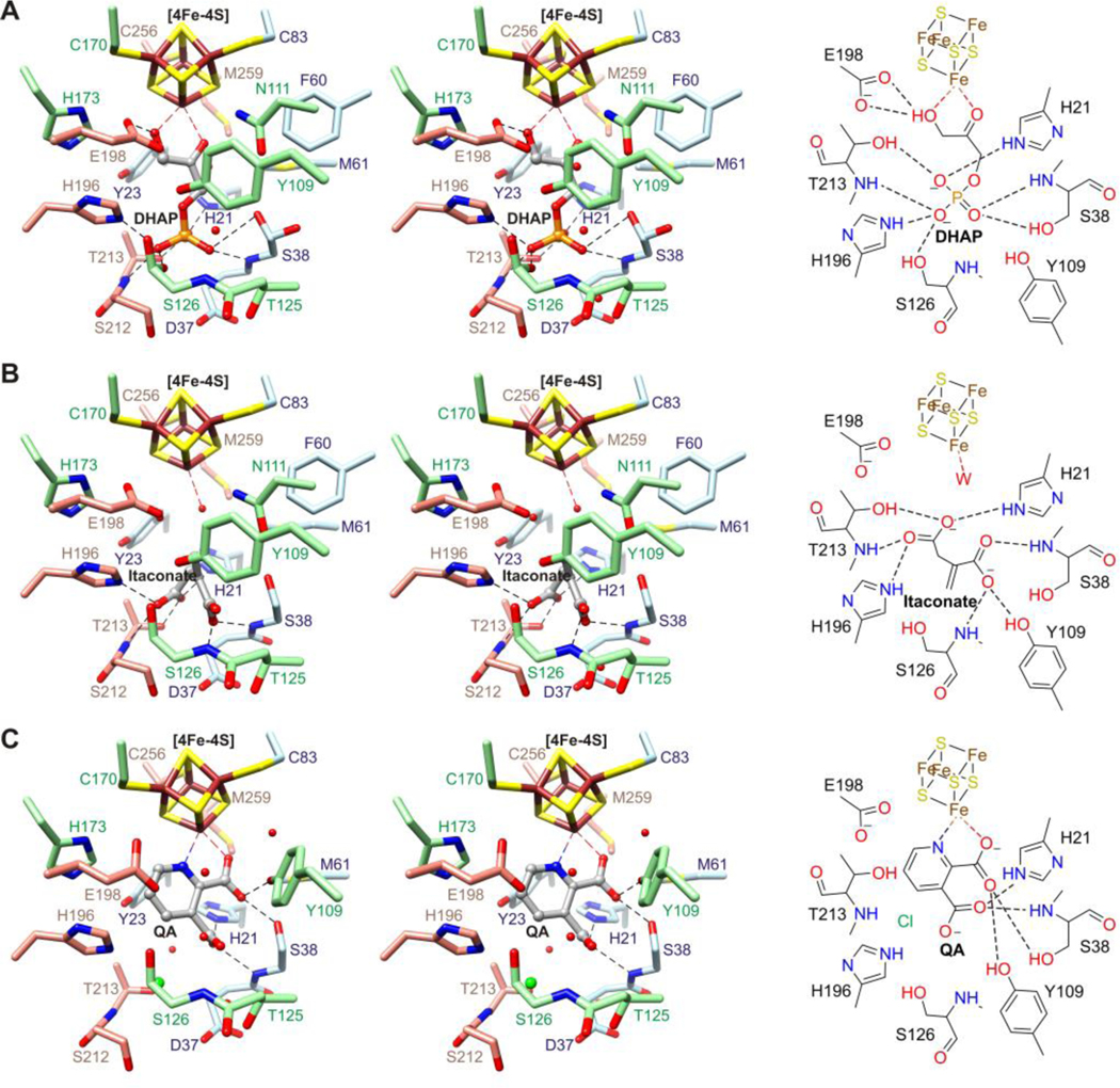
Stereo diagrams (left) and schematic drawings (right) of the active site of PhNadA with bound (A) DHAP, (B) itaconate, and (C) QA. Black broken lines denote potential hydrogen bonds, red spheres denote waters, and the green sphere denotes chloride.
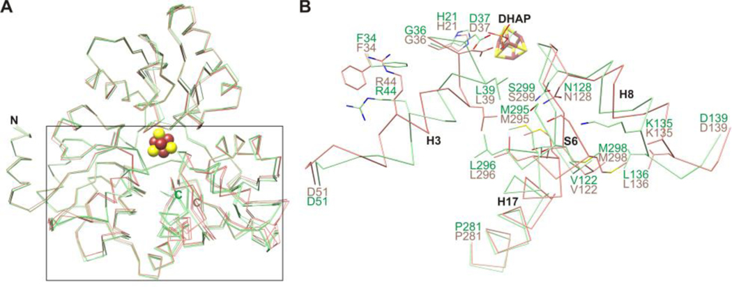
(A) Superimposition of Cα traces showing open (holo and L-malate bound structures; salmon) and closed (DHAP, itaconate, maleate, and citraconate bound structures; light green) states of PhNadA. (B) Close-up of holo and DHAP bound structures indicating side chain clashes.
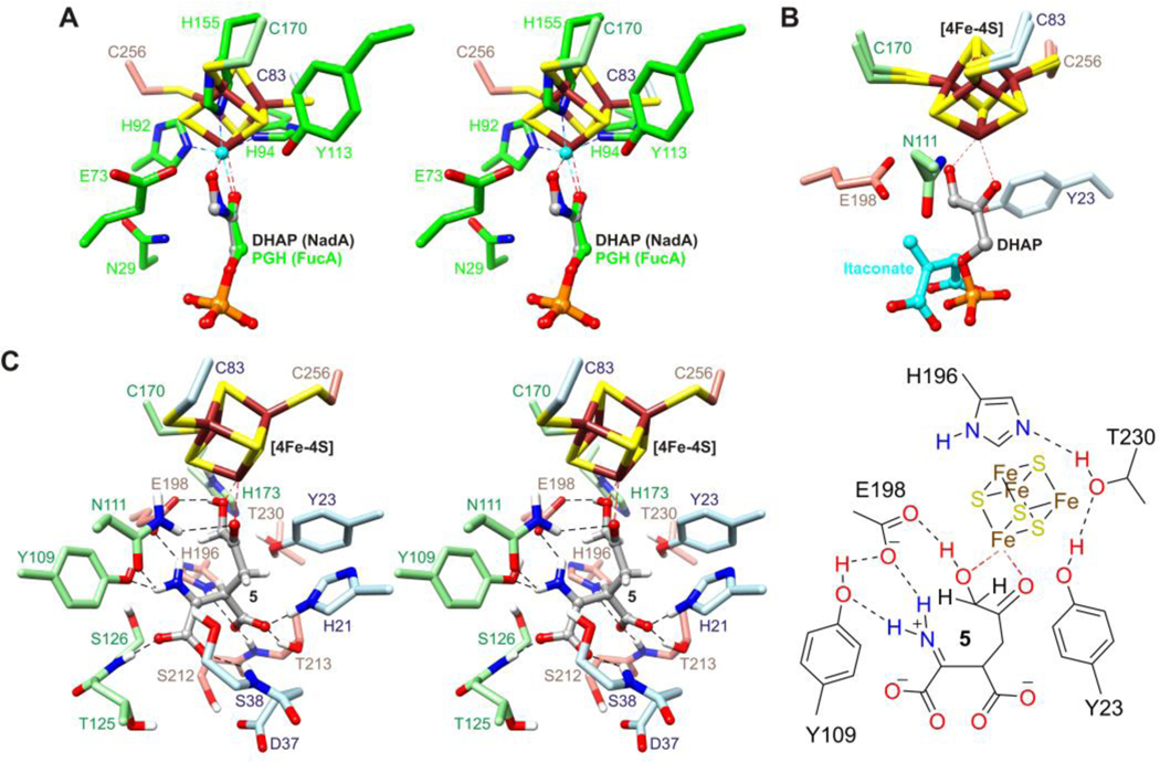
(A) Stereo diagram of the superimposition of DHAP bound to the [4Fe-4S] cluster in PhNadA onto PGH bound to zinc (cyan) in FucA (green). (B) Superimposition of the [4Fe-4S] cluster with bound DHAP onto the [4Fe-4S] cluster in the itaconate bound structure of PhNadA. (C) Stereo diagram (left) and schematic drawing (right) of intermediate 5 based on the result in panel B, with C1 and Cβ joined, followed by energy minimization with positional restraints applied to the protein atoms.
Similar articles
-
An Unexpected Species Determined by X-ray Crystallography that May Represent an Intermediate in the Reaction Catalyzed by Quinolinate Synthase.J Am Chem Soc. 2019 Sep 11;141(36):14142-14151. doi: 10.1021/jacs.9b02513. Epub 2019 Aug 26. J Am Chem Soc. 2019. PMID: 31390192 Free PMC article.
-
Structure of Quinolinate Synthase from Pyrococcus horikoshii in the Presence of Its Product, Quinolinic Acid.J Am Chem Soc. 2016 Jun 15;138(23):7224-7. doi: 10.1021/jacs.6b02708. Epub 2016 Jun 2. J Am Chem Soc. 2016. PMID: 27224840
-
Characterization of quinolinate synthases from Escherichia coli, Mycobacterium tuberculosis, and Pyrococcus horikoshii indicates that [4Fe-4S] clusters are common cofactors throughout this class of enzymes.Biochemistry. 2008 Oct 14;47(41):10999-1012. doi: 10.1021/bi801268f. Epub 2008 Sep 20. Biochemistry. 2008. PMID: 18803397 Free PMC article.
-
DHAP-dependent aldolases from (hyper)thermophiles: biochemistry and applications.Extremophiles. 2014 Jan;18(1):1-13. doi: 10.1007/s00792-013-0593-x. Epub 2013 Oct 29. Extremophiles. 2014. PMID: 24166576 Review.
-
Quinolinate Synthase: An Example of the Roles of the Second and Outer Coordination Spheres in Enzyme Catalysis.Chem Rev. 2022 Jul 27;122(14):12110-12131. doi: 10.1021/acs.chemrev.1c00869. Epub 2022 May 10. Chem Rev. 2022. PMID: 35536891 Review.
Cited by
-
An Unexpected Species Determined by X-ray Crystallography that May Represent an Intermediate in the Reaction Catalyzed by Quinolinate Synthase.J Am Chem Soc. 2019 Sep 11;141(36):14142-14151. doi: 10.1021/jacs.9b02513. Epub 2019 Aug 26. J Am Chem Soc. 2019. PMID: 31390192 Free PMC article.
-
Structure-based insights into the mechanism of [4Fe-4S]-dependent sulfur insertase LarE.Protein Sci. 2024 Feb;33(2):e4874. doi: 10.1002/pro.4874. Protein Sci. 2024. PMID: 38100250 Free PMC article.
References
-
- Begley TP, Kinsland C, Mehl RA, Osterman A, and Dorrestein P. (2001), Vitam. Horm 61, 103–119. - PubMed
-
- Nasu S, Wicks FD, and Gholson RK (1982), J. Biol. Chem 257, 626–632. - PubMed
-
- Cicchillo RM, Tu L, Stromberg JA, Hoffart LM, Krebs C, and Booker SJ (2005), J. Am. Chem. Soc 127, 7310–7311. - PubMed
-
- Gardner PR, and Fridovich I. (1991), Arch. Biochem. Biophys 284, 106–111. - PubMed
Publication types
MeSH terms
Substances
Grants and funding
LinkOut - more resources
Full Text Sources
Other Literature Sources
