

Case control study: magnetic resonance spectroscopy of brain in HIV infected patients
- PMID: 27405321
- PMCID: PMC4942893
- DOI: 10.1186/s12883-016-0628-x
Case control study: magnetic resonance spectroscopy of brain in HIV infected patients
Abstract
Background: In vivo proton magnetic resonance spectroscopy ((1)H-MRS) studies on brain in HIV infected patients have shown significant alteration in neuro-biochemicals.
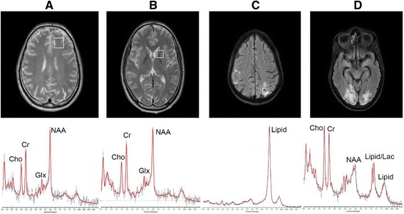
Methods: In this study, we measured the neuro-biochemical metabolites from the left frontal white matter (FWM) and left basal ganglia (BG) caudate head nucleus in 71 subjects that include 30 healthy controls, 20 asymptomatic HIV and 21 HIV patients with CNS lesion. Proton MR spectra were acquired at 3 T MRI system and the concentration (institutional units) of tNAA (N-acetylaspartate, NAA + N-acetylaspartylglutamate, NAAG), tCr (Creatine, Cr + phosphocreatine, PCr), choline containing compounds (tCho), glutamate + glutamine (Glx) and lipid and macromolecules at 0.9 ppm were determined using LC Model.
Results: In BG, the concentration of tNAA (6.71 ± 0.64) was decreased and in FWM, the concentration of Glx (20.4 ± 7.8), tCr (9.14 ± 3.04) and lipid and macromolecules at 0.9 ppm (8.69 ± 2.96) were increased in HIV patients with CNS lesion. In healthy controls, the concentration of tNAA in BG was 7.31 ± 0.47 and concentration of Glx, tCr and lipid and macromolecules in FWM were 15.0 ± 6.06, 6.95 ± 2.56, 5.59 ± 1.56, respectively.
Conclusion: Reduced tNAA in BG suggests neuronal loss in HIV patients with CNS lesion while increased Glx in FWM may suggest excito-toxicity. In addition, increased levels of tCr in FWM of HIV patients were observed. The study indicates region specific metabolic changes in tNAA, tCr and Glx in brain of HIV infected patients.
Keywords: AIDS; Glutamate; HIV; MRS; Magnetic resonance spectroscopy.
Figures
References
-
- Global summary of the AIDS epidemic. http://www.who.int/hiv/data/epi_core_july2015.png?ua=1. Accessed 9 July 2016.
-
- Annual Report. In: Control DoA, editor. New Delhi: Ministry of Health and Family Welfare; 2013–2014. www.mohfw.nic.in/index1.
-
- Grant I, Atkinson JH, Hesselink JR, Kennedy CJ, Richman DD, Spector SA, McCutchan JA. Evidence for early central nervous system involvement in the acquired immunodeficiency syndrome (AIDS) and other human immunodeficiency virus (HIV) infections: Studies with neuropsychologic testing and magnetic resonance imaging. Ann Intern Med. 1987;107(6):828–36. doi: 10.7326/0003-4819-107-6-828. - DOI - PubMed
-
- Fauci AS, Lane HC. HIV neurology. In Hauser SI and Josephson (Eds.), Harrison’s neurology in clinical medicine (2nd ed.). New York: McGraw-Hill; 2010.
-
- Chong W, Sweeney B, Wilkinson I, Paley M, Hall-Craggs M, Kendall B, Shepard J, Beecham M, Miller R, Weller I. Proton spectroscopy of the brain in HIV infection: correlation with clinical, immunologic, and MR imaging findings. Radiology. 1993;188(1):119–24. doi: 10.1148/radiology.188.1.8099750. - DOI - PubMed
MeSH terms
Substances
LinkOut - more resources
Full Text Sources
Other Literature Sources
Medical
