. 2016 Jul 13:6:29784.
doi: 10.1038/srep29784.

Function of human pluripotent stem cell-derived photoreceptor progenitors in blind mice

Affiliations

Function of human pluripotent stem cell-derived photoreceptor progenitors in blind mice

Alona O Barnea-Cramer et al. Sci Rep. .

Abstract

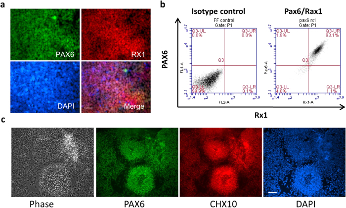
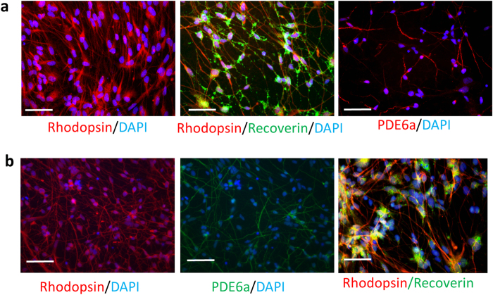
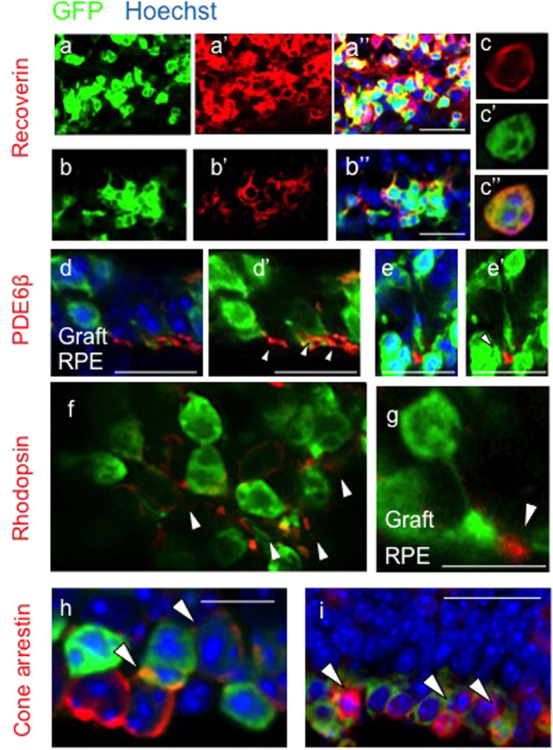
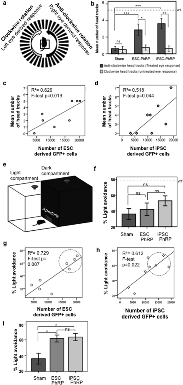
Photoreceptor degeneration due to retinitis pigmentosa (RP) is a primary cause of inherited retinal blindness. Photoreceptor cell-replacement may hold the potential for repair in a completely degenerate retina by reinstating light sensitive cells to form connections that relay information to downstream retinal layers. This study assessed the therapeutic potential of photoreceptor progenitors derived from human embryonic and induced pluripotent stem cells (ESCs and iPSCs) using a protocol that is suitable for future clinical trials. ESCs and iPSCs were cultured in four specific stages under defined conditions, resulting in generation of a near-homogeneous population of photoreceptor-like progenitors. Following transplantation into mice with end-stage retinal degeneration, these cells differentiated into photoreceptors and formed a cell layer connected with host retinal neurons. Visual function was partially restored in treated animals, as evidenced by two visual behavioral tests. Furthermore, the magnitude of functional improvement was positively correlated with the number of engrafted cells. Similar efficacy was observed using either ESCs or iPSCs as source material. These data validate the potential of human pluripotent stem cells for photoreceptor replacement therapies aimed at photoreceptor regeneration in retinal disease.


Conflict of interest statement

W.W., S.-J.L., C.L., H.H. and R.L. are employees of Astellas Institute for Regenerative Medicine, a biotechnology company in the field of stem cells and regenerative medicine.

Figures

Figure 1
Figure 1. In vitro Differentiation of Human Embryonic Stem Cells towards Retinal Neural Progenitors.
(a) Immunofluorescence staining shows co-expression of PAX6 and RX1 on day 13 eye field progenitors. (b) Quantification of PAX6 and RX1double positive eye field progenitors by flow cytometry analysis which shows >90% of them expressing both PAX6 and RX1 proteins. (c) Phase contrast image shows neural rosette structures of retinal neuronal progenitor cells (RNPC, far left) and immunofluorescence staining shows expression of PAX6 and CHX10 on RNPC at about day 30 after initial differentiation in vitro. Scale bar, 50 μm.
Figure 2
Figure 2. In vitro Differentiation of Retinal Neural Progenitors towards Photoreceptor-like Progenitors.
(a) Immunofluorescence staining shows the expression of transcription factors NRL, NR2E3 and CRX in PhRPs at 90 –100 days after in vitro differentiation. CHX10 and neuN genes are negative in PhRPs at this stage; the upper right corner of the CHX10 image shows positive expression of CHX10 in RNPCs at day 30 (positive control); the upper right corner of the neuN image shows positive expression of neuN in mouse central nerve cells (positive control). 2nd antibody only also shows negative staining. (b) Quatification of intracellular staining of NRL, NR2E3 and CRX as determined by Flow Cytometry analyses. Scale bar, 50 μm.
Figure 3
Figure 3. In vitro Generation of Mature Photoreceptor-like Cells from Human ESC/iPS-Derived PhRPs.
Expression of rod photoreceptor markers, rhodopsin, recoverin and PDE6α in hESC (a) and iPSC-derived (b) photoreceptor-like cells two week after in vitro maturation. Scale bar, 50 μm.
Figure 4
Figure 4. Transplanted Human ESC-PhRPs and iPSC-PhRPs Survive in the Subretinal Space of rd1 Mice.
Scanning laser ophthalmoscopy (SLO) was performed in vivo three weeks post transplantation to assess the extent of surviving donor cells; GFP positive cells are observed in autofluorescence (AF) mode as white dots or clusters (black areas represent areas of retina which were not seeded with transplanted cells, due to incomplete detachment of the retina around the optic nerve head). Representative near-infrared (NIR) and AF fundus images of rd1 mice show a homogeneous presence of GFP+ cells in the two treatment groups: ESC-PhRPs (a) and iPSC-PhRPs (b). Histological assessment 3 weeks post transplantation revealed ESC-PhRP (c-c’) and iPSC-PhRP (d-d’) derived cell layers (green) between the retinal pigment epithelium (RPE) and inner nuclear layer (INL) of the rd1 retina, replacing the absent outer nuclear layer (ONL) in the adult rd1 mice; (e-e’) GFP+ cells were stained with human nuclear antigen (HNA) which co-localized with GFP; indicating that the GFP signal observed in vivo in treated animals was indeed an indicator of transplanted human PhRPs. Scale bar, 25 μm.
Figure 5
Figure 5. Transplanted Human ESC-PhRPs and iPSC-PhRPs Express Mature Photoreceptor Markers in vivo.
Immunofluorescence staining 3 weeks post transplantation shows expression of mature photoreceptor markers in transplanted human PhRPs (green). In all images cells are located in the subretinal space and oriented so that the host INL is located at the top of the image and the RPE at the bottom. The pan-photoreceptor marker recoverin was observed within the reconstructed layer of cells in animals treated with both ESC-PhRPs (a) and iPSC-PhRPs (b,c). The rod specific enzyme phosphodiesterase β6 (PDE6b), which is necessary in phototransduction and is absent in rd1 mice due to mutation was reinstated in the retina and located in the outer processes of transplanted ESC-PhRPs (d-d’) and iPSC-PhRPs (e-e’). The rod specific protein rhodopsin, which is normally located in outer segment membrane disk was also observed in outer segments of ESC-PhRPs (f) and iPSC-PhRPs (g). Cone arrestin was observed in GFP+ cells, indicating that a subset of human cells matured to produce cone photoreceptors. Scale bar, 20 μm.
Figure 6
Figure 6. Graft-Host Connectivity.
(a-a”’) Expression of synaptophysin, a synaptic marker, was present between the host INL and the GFP-positive graft. Synaptophysin is localized between the host and graft and is expressed in transplanted cells (white arrows) indicating synaptic transmission between human iPSC-derived grafted cells and the host rd1 retina; (a) GFP (human cells); (a’) synaptophysin; (a”) merged image of GFP and synaptophysin; and (a”’) merged image of GFP, synaptophysin and DAPI. The dashed line delineates the boundary between the host INL and the graft. (b-b”’) Glial fibrillary acidic protein (GFAP), a protein expressed by inner retinal astrocytes and activated Müller glia, is expressed by the glial cells of the host retina (red). Gliosis in the degenerate retina may occur to protect the retina from further damage, and a horizontal glial scar at the edge of the host ONL was observed in some areas of the retina (black arrow). However, glial processes were also observed to extend into the graft (green), without formation of a complete glial barrier between host and graft (white arrows). (b) GFP (human cells); (b’) GFAP+ host glial cells; (b”) merged image of ((b) GFP) and ((b’) GFAP); (b”’) merged image of GFP, GFAP and DAPI. Scale bar, 20 μm.
Figure 7
Figure 7. Recovery of Basic Visual Responses in rd1 Mice Following Transplantation of Human PhRPs Correlates to Number of Engrafted Cells.
(a) Schematic of the optomotor response (OMR) test arena and expected response to the direction of drum rotation. (b) Mean OMR 3 weeks post-transplantation indicating an improvement in OMR driven by treated eyes (dark grey) compared to paired untreated eyes (light grey) after transplantation of ESC-PhRPs (paired sample t-test, t = 2.86, p = 0.024) and iPSC-PhRPs (paired sample t-test, t = 5.02, p = 0.002); In the sham treated group there were no differences in OMR driven by treated and untreated eyes (paired sample t test, t = 0.31, ns). Furthermore, OMR was improved in PhRP treatment groups compared to sham treatment (one way-ANOVA, F = 7.8, p = 0.003), with an increase in the response in both ESC-PhRP (p < 0.05) and iPSC-PhRP (p < 0.005) treated animals (Bonferroni test for multiple comparisons). (c) A positive correlation was observed between number of head tracks and number of GFP+ cells in animals treated with ESC-PhRPs (n = 8, R2 = 0.729, F = 16.13, p < 0.01) and (d) iPSC-PhRPs (n = 8, R2 = 0.612, F = 9.46, p < 0.05). (*p < 0.5, **p < 0.01). (e) Schematic of the light avoidance apparatus. (f) There were no differences between the three groups in mean light avoidance responses (F = 1.43, p = 0.261 [ns]). However, a positive correlation was observed between number of GFP+ cells and light avoidance behavior in individual animals of (g) ESC-PhRP (R2 = 0.729, F = 16.13, p < 0.01) and (h) iPSC-PhRP treated group (R2 = 0.612, F = 9.46, p < 0.05). (i) Comparing only animals with above-median numbers of GFP+ cells (encircled in (g,h)) a significant difference was observed between the three groups (X2 = 6 (df2), p < 0.05) showing improvement in ESC-PhRP treated (n = 4, p < 0.05) and iPSC-PhRP treated (n = 4, p < 0.05) subgroups. The dashed line in (b,f) represents the mean response of age-matched wild-type mice. Error bars represent ± S.E.M.

References

    1. Hartong D. T., Berson E. L. & Dryja T. P. Retinitis pigmentosa. Lancet 368, 1795–1809, 10.1016/s0140-6736(06)69740-7 (2006). - DOI - PubMed
    1. Barber A. C. et al.. Repair of the degenerate retina by photoreceptor transplantation. Proceedings of the National Academy of Sciences of the United States of America 110, 354–359, 10.1073/pnas.1212677110 (2013). - DOI - PMC - PubMed
    1. Luo J. et al.. Human retinal progenitor cell transplantation preserves vision. The Journal of biological chemistry 289, 6362–6371, 10.1074/jbc.M113.513713 (2014). - DOI - PMC - PubMed
    1. Singh M. S. et al.. Reversal of end-stage retinal degeneration and restoration of visual function by photoreceptor transplantation. Proceedings of the National Academy of Sciences of the United States of America 110, 1101–1106, 10.1073/pnas.1119416110 (2013). - DOI - PMC - PubMed
    1. Pearson R. A. et al.. Restoration of vision after transplantation of photoreceptors. Nature 485, 99–103, 10.1038/nature10997 (2012). - DOI - PMC - PubMed

Publication types

MeSH terms