. 2016 Aug 26;291(35):18465-73.
doi: 10.1074/jbc.M116.727750. Epub 2016 Jul 12.

Controlling PTEN (Phosphatase and Tensin Homolog) Stability: A DOMINANT ROLE FOR LYSINE 66

Affiliations

Controlling PTEN (Phosphatase and Tensin Homolog) Stability: A DOMINANT ROLE FOR LYSINE 66

Amit Gupta et al. J Biol Chem. .

Abstract

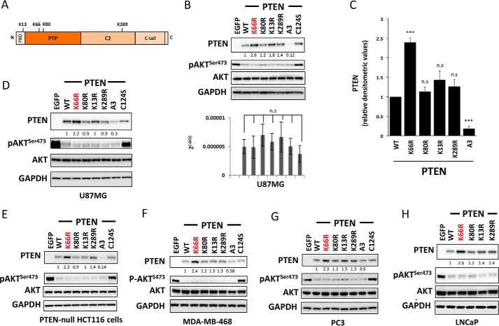
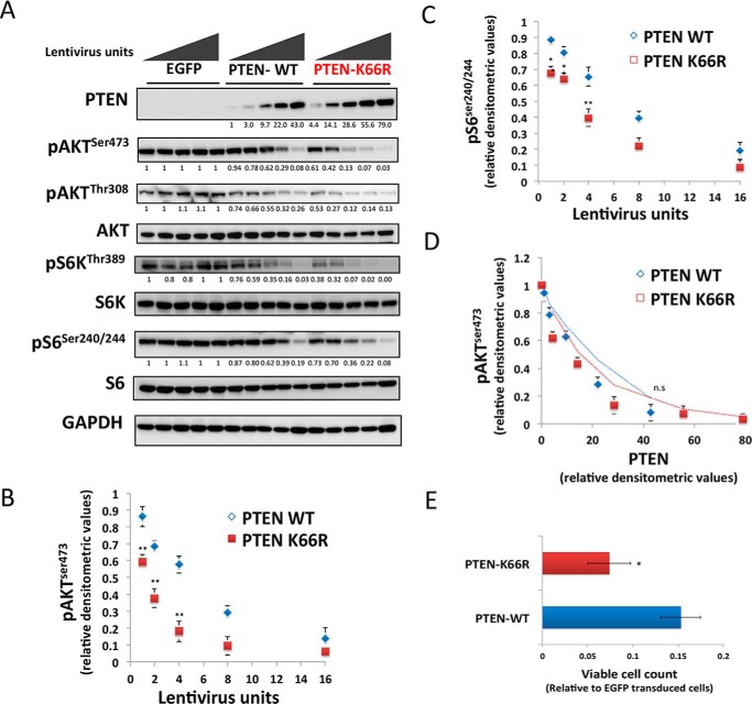
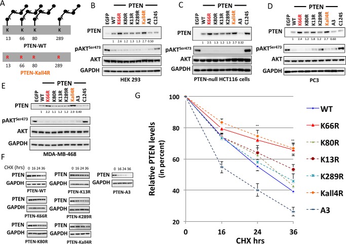
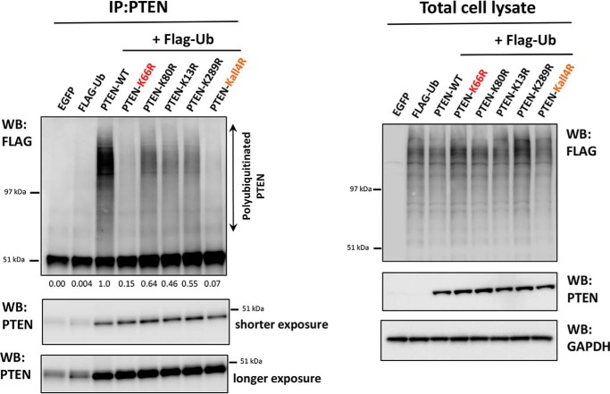
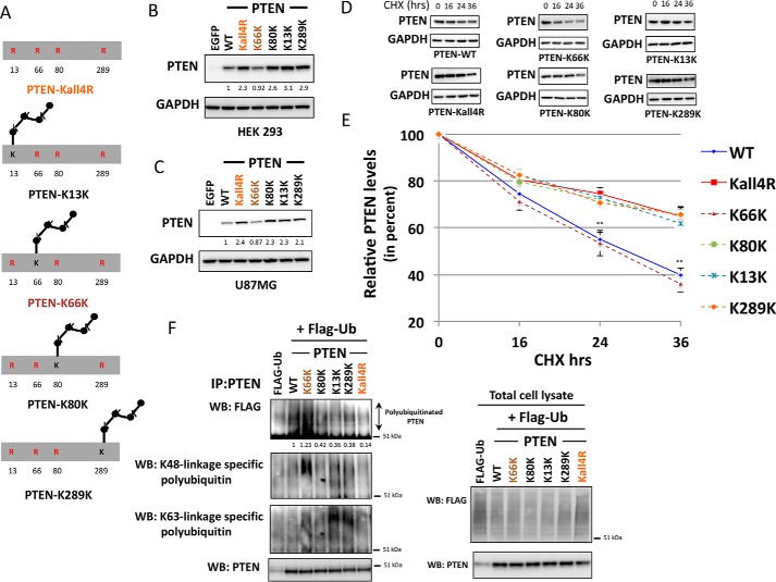
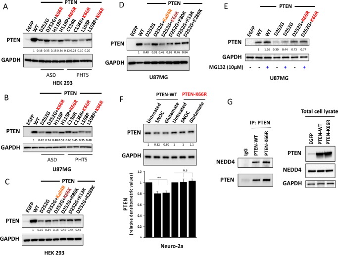
Phosphatase and tensin homolog (PTEN) is a phosphoinositide lipid phosphatase and one of the most frequently disrupted tumor suppressors in many forms of cancer, with even small reductions in the expression levels of PTEN promoting cancer development. Although the post-translational ubiquitination of PTEN can control its stability, activity, and localization, a detailed understanding of how PTEN ubiquitination integrates with other cellular regulatory processes and may be dysregulated in cancer has been hampered by a poor understanding of the significance of ubiquitination at individual sites. Here we show that Lys(66) is not required for cellular activity, yet dominates over other PTEN ubiquitination sites in the regulation of protein stability. Notably, combined mutation of other sites (Lys(13), Lys(80), and Lys(289)) has relatively little effect on protein expression, protein stability, or PTEN polyubiquitination. The present work identifies a key role for Lys(66) in the regulation of PTEN expression and provides both an opportunity to improve the stability of PTEN as a protein therapy and a mechanistic basis for efforts to stabilize endogenous PTEN.

Keywords: cancer; phosphatase; phosphatase and tensin homolog (PTEN); phosphoinositide; phosphoinositide 3-kinase; protein stability; tumor suppressor; ubiquitylation (ubiquitination).


Figures

FIGURE 1.
FIGURE 1.
Mutation of PTEN lysine 66 increases PTEN expression. A, a schematic illustrating the 403-amino acid PTEN protein and the ubiquitinated lysine residues analyzed in this study. B and C, PTEN-null U87MG glioblastoma cells were transduced with similar units of lentivirus particles encoding for PTEN WT or the indicated PTEN mutants. Control cells were transduced with viruses encoding EGFP. PTEN expression and AKT phosphorylation were investigated by Western blotting of total cell lysates using total and phospho-specific antibodies. GAPDH levels were used as a loading control. A representative blot from at least three independent experiments is shown. Parallel samples from U87MG cells transduced with PTEN WT or mutants were used to analyze the levels of PTEN mRNA by quantitative PCR (B, lower panel). The PTEN transcript levels are expressed relative to GAPDH. y axis represents the average levels of transcript ± S.E. from three experiments each performed in duplicate. No significant difference in the transcript levels was observed between PTEN WT and each PTEN mutant-expressing sample. C, bars representing the relative densitometric values of PTEN expression. The quantitation is derived from three independent experiments and values are mean ± S.E. PTEN K66R was significantly more stable compared with PTEN WT (p value < 0.001). No significant changes in PTEN levels were observed with mutation of other ubiquitin sites (n.s., non significant). D, PTEN-null U87MG glioblastoma cells were transfected with plasmid expression vectors encoding for PTEN WT, PTEN mutants, or EGFP as a control. Cells were lysed 48 h post-transfection and PTEN expression and AKT phosphorylation were investigated by Western blotting analysis of total cell lysates using total and phospho-specific antibodies. GAPDH levels were used as a loading control. A representative blot from three independent experiments is shown. These plasmid transfection experiments recapitulate the increase in PTEN expression with K66R mutation relative to PTEN WT (p value < 0.001) as observed with a lentiviral transduction approach. E–H, PTEN-null clone of HCT116 colon cancer cells (E), MDA-MB-468 breast cancer cells (F), PC3 (G), and LNCaP prostate cancer cells (H) were transduced with similar units of lentivirus particles encoding for PTEN WT or the indicated PTEN mutants. Control cells were transduced with viruses encoding EGFP. PTEN expression and AKT phosphorylation were investigated by Western blotting analysis of total cell lysates using total and phosphospecific antibodies. GAPDH levels were used as a loading control. In each case a representative blot from at least three independent experiments is shown. In all cases, as in U87MG cells, expression of PTEN K66R was significantly higher than wild-type (p value < 0.01).
FIGURE 2.
FIGURE 2.
Mutation of PTEN lysine 66 increases PTEN expression and shows a concomitant greater effect on downstream cell signaling and proliferation. A–D, U87MG cells were transduced with increasing doses of lentiviruses encoding EGFP, PTEN WT, or PTEN K66R (A) PTEN expression and the effect on downstream cell signaling was analyzed by immunoblotting with PTEN, phospho-AKT Ser473, phospho-AKT Thr308, AKT, phospho-S6K Thr389, S6K, phospho-S6 Ser240/244, and S6 antibodies. GAPDH levels were used as a loading control. B and C, graph representing the effect of increasing doses of PTEN WT or PTEN K66R lentiviruses on the suppression of (B) AKT Ser473 and (C) S6 Ser240/244 cellular phosphorylation, relative to EGFP-transduced cells. y axis shows the mean relative densitometric values of phosphorylation ± S.E. from three independent experiments. x axis shows the units of lentivirus used. PTEN K66R lentiviruses were significantly more effective in suppressing both AKT and S6 phosphorylation compared with similar dose of PTEN WT lentiviruses. D, the graph representing the specific activity of PTEN-WT versus PTEN K66R. The average relative densitometric values of AKT Ser473 phosphorylation from three independent experiments for PTEN WT and PTEN K66R was plotted against the relative PTEN expression levels with increasing doses of PTEN WT or PTEN K66R lentiviruses. E, PTEN-null U87MG cells were transduced with similar units of lentiviruses encoding EGFP, PTEN WT, or PTEN K66R and the viable cell number was quantified bye 5 days post-transduction using a 3-(4,5-dimethylthiazol-2-yl)-2,5-diphenyltetrazolium bromide assay. x axis shows the mean measured cellular metabolic activity relative to EGFP-transduced cells ± S.E. from three experiments each performed in triplicate. t test: **, p value < 0.01; *, p value < 0.05; n.s., non significant.
FIGURE 3.
FIGURE 3.
Dominant role of PTEN lysine 66 ubiquitination site in the regulation of PTEN expression and stability. A, a schematic illustrating the mutant PTEN Kall4R with all the four studied potential ubiquitination sites (lysine 13, 66, 80, and 289) mutated to arginine (Arg). B–E, HEK293T cells (B), PTEN-null HCT116 colon cancer cells (C), PC3 prostate cancer cells (D), and MDA-MB-468 breast cancer cells (E) were transduced with similar units of lentivirus particles encoding for PTEN WT, the indicated PTEN mutants, or control EGFP. PTEN expression and AKT phosphorylation were investigated by Western blotting analysis of cell lysates using total and phospho-specific antibodies. GAPDH levels were used as a loading control. A representative blot from three independent experiments is shown. F and G, the stability of PTEN ubiquitin-site mutants was determined using protein synthesis inhibitor cycloheximide (CHX). F, PTEN-null U87MG cells transduced with viruses encoding PTEN WT or PTEN mutants were treated with 100 μg/ml of CHX for the indicated times, followed by immunoblot analysis with anti-PTEN or anti-GAPDH antibodies. The blots shown are representative of three independent experiments. G, plot showing densitometric analysis of the CHX assay. y axis shows the relative PTEN levels (in percent) after normalizing with GAPDH levels. The quantitation is derived from three independent experiments and values are mean ± S.E. Both PTEN K66R and PTEN Kall4R, with Lys66 mutation, were significantly more stable compared with PTEN WT (p value < 0.01).
FIGURE 4.
FIGURE 4.
Dominant role of the PTEN lysine 66 ubiquitination site in the regulation of PTEN polyubiquitination. Cellular assay for PTEN ubiquitination. U87MG cells were transfected with an expression vector encoding FLAG-ubiquitin (FLAG-Ub). 6 h after transfection cells were transduced with viruses encoding PTEN WT or PTEN mutants. Lentiviruses were titrated prior to the experiment to ensure similar levels of PTEN protein in each sample. 48 h post-transfection cells were treated with 10 μm MG132 for 5 h. Cells were lysed, and PTEN was immunoprecipitated (IP) before Western blotting (WB) of these immunoprecipitates with antibodies raised against the FLAG epitope and PTEN. In parallel, total cell lysates were immunoblotted with antibodies raised against the FLAG epitope, PTEN, or GAPDH (left panel).
FIGURE 5.
FIGURE 5.
Contribution of individual ubiquitination sites in regulating PTEN expression, stability, and PTEN polyubiquitination. A, schematic illustrating PTEN mutants generated to assess the contribution of individual ubiquitination sites to expression and stability. B and C, HEK293T cells (B) and PTEN-null U87MG glioblastoma cells (C) were transduced with similar units of lentivirus particles encoding for PTEN WT or PTEN mutants or EGFP. PTEN expression was investigated by Western blotting (WB) analysis, GAPDH levels were used as a loading control. A representative blot from three independent experiments is shown. D and E, the stability of these PTEN mutants was determined using protein synthesis inhibitor cycloheximide (CHX). D, U87MG cells transduced with viruses encoding PTEN WT or PTEN mutants were treated with 100 μg/ml of CHX for the indicated times, followed by immunoblot analysis with anti-PTEN or anti-GAPDH antibodies. The blots shown are representative of three independent experiments. E, plot showing densitometric analysis of the CHX assay. y axis shows the relative percentage PTEN levels after normalizing to GAPDH. The quantitation is derived from three independent experiments and values are mean ± S.E. PTEN K66K, with only Lys66 ubiquitination site available, showed significantly lower stability than PTEN Kall4R (p value < 0.01). PTEN mutants that included the K66R change (i.e. PTEN Kall4R, K13K, K80K, and K289K) showed more or less similar stability with data points falling on top of each other. F, cellular assay for PTEN ubiquitination. U87MG cells were transfected to express FLAG-ubiquitin (FLAG-Ub) and 6 h later transduced with viruses encoding PTEN WT or the indicated PTEN mutants. Lentiviruses were titrated prior to the experiment to ensure similar levels of PTEN protein is expressed in each sample. 48 h post-transfection, cells were treated with 10 μm MG132 for 5 h. Cells were lysed, and PTEN was immunoprecipitated (IP) before Western blotting analysis of these immunoprecipitates with antibodies raised against the FLAG epitope, Lys48 linkage-specific polyubiquitin, Lys63 linkage-specific polyubiquitin, and PTEN. A smear of high molecular weight in the FLAG Western blot represents polyubiquitinated PTEN. A stronger signal is observed with a Lys48 linkage-specific antibody in PTEN K66K mutant that has only the Lys66 site available for ubiquitination out of the four studied ubiquitination sites. In parallel, total cell lysates were immunoblotted with antibodies raised against the FLAG epitope, PTEN, or GAPDH (F, lower panel).
FIGURE 6.
FIGURE 6.
Mutating lysine 66 counteracts destabilizing PTEN mutations associated with autism and tumor syndromes and NO-mediated proteasomal degradation. HEK293T cells (A) and PTEN-null U87MG cells (B) were transfected with vectors encoding PTEN WT or unstable PTEN mutants associated with autism spectrum disorder (ASD) and tumor syndromes (PHTS) or the same unstable mutants carrying an additional K66R mutation. Cells were lysed and PTEN expression was investigated by Western blotting analysis. GAPDH levels were used as a loading control. Representative blots from three independent experiments is shown. Additional mutation of K66R significantly increased the protein expression level of the unstable PTEN mutant proteins D252G, H118P (p value < 0.001), and L108P (p value < 0.01). C and D, effects of PTEN ubiquitination site mutation in PTEN D252G mutant background. HEK293T cells (C) and U87MG cells (D) were transfected as indicated with expressing vectors encoding PTEN WT or PTEN mutants. Cells were lysed and PTEN expression was investigated by Western blotting analysis. GAPDH levels were used as a loading control. A representative blot from three independent experiments is shown. All mutant PTEN proteins containing the K66R mutation showed significant stabilization of D252G mutant enzyme (p value < 0.01). Mutant PTEN D252G-K66K retaining Lys66 in its wild-type form showed no significant effect on D252G mutant enzyme stability. E, U87MG cells transfected with the indicated expression vectors encoding PTEN WT or PTEN mutants were treated for 5 h with 10 μm MG132 proteasome inhibitor before PTEN expression was analyzed by Western blotting analysis for PTEN and GAPDH. F, mouse neuroblastoma Neuro-2a cells were transfected with quantities of the indicated expression vectors encoding PTEN WT or PTEN K66R engineered to lead to similar expression of the two proteins. Cells were treated for 4 h with 200 μm freshly prepared S-nitrosocysteine (SNOC) or 200 μm glutamate before PTEN expression was analyzed by Western blotting for PTEN and GAPDH. A representative blot from three independent experiments is shown (F, lower panel). Bar representing relative densitometric values of PTEN expression after SNOC and glutamate treatments in PTEN-WT and PTEN K66R-transfected cells. The quantitation is derived from three independent experiments and values are mean ± S.E. SNOC acts as a physiological NO donor and glutamate is neurotoxic compound, which induces NO generation. SNOC and glutamate-mediated S-nitrosylation of PTEN has been shown to lead to enhanced PTEN ubiquitination leading to its proteasome-mediated degradation (31). A modest but significant decrease in PTEN levels is observed with SNOC and glutamate treatment in PTEN WT overexpressing cells (two-tailed t test, p value < 0.01). PTEN-K66R does not show this decrease in PTEN levels. G, co-immunoprecipitation of PTEN and NEDD4 in PTEN-WT and PTEN K66R-transfected 293T cells. Quantities of expression vectors encoding PTEN WT or PTEN K66R were engineered to lead to similar expression of the two proteins. PTEN was immunoprecipitated (IP) and PTEN-bound endogenous NEDD4 was immunoblotted.

Similar articles

Cited by

References

    1. Leslie N. R., and Foti M. (2011) Non-genomic loss of PTEN function in cancer: not in my genes. Trends Pharmacol. Sci. 32, 131–140 - PubMed
    1. Song M. S., Salmena L., and Pandolfi P. P. (2012) The functions and regulation of the PTEN tumour suppressor. Nat. Rev. Mol. Cell Biol. 13, 283–296 - PubMed
    1. Worby C. A., and Dixon J. E. (2014) PTEN. Annu. Rev. Biochem. 83, 641–669 - PubMed
    1. Hopkins B. D., Fine B., Steinbach N., Dendy M., Rapp Z., Shaw J., Pappas K., Yu J. S., Hodakoski C., Mense S., Klein J., Pegno S., Sulis M. L., Goldstein H., Amendolara B., et al. (2013) A secreted PTEN phosphatase that enters cells to alter signaling and survival. Science 341, 399–402 - PMC - PubMed
    1. Liang H., He S., Yang J., Jia X., Wang P., Chen X., Zhang Z., Zou X., McNutt M. A., Shen W. H., and Yin Y. (2014) PTENα, a PTEN isoform translated through alternative initiation, regulates mitochondrial function and energy metabolism. Cell Metab. 19, 836–848 - PMC - PubMed

Publication types

LinkOut - more resources