. 2016 Jul 13:7:12197.
doi: 10.1038/ncomms12197.

BMP and retinoic acid regulate anterior-posterior patterning of the non-axial mesoderm across the dorsal-ventral axis

Affiliations

BMP and retinoic acid regulate anterior-posterior patterning of the non-axial mesoderm across the dorsal-ventral axis

Richard W Naylor et al. Nat Commun. .

Abstract

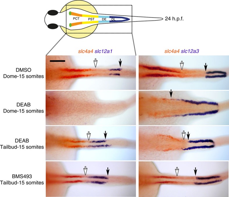
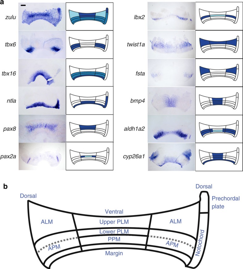
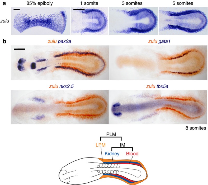
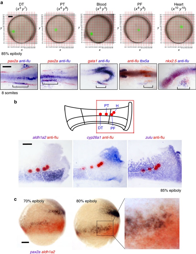
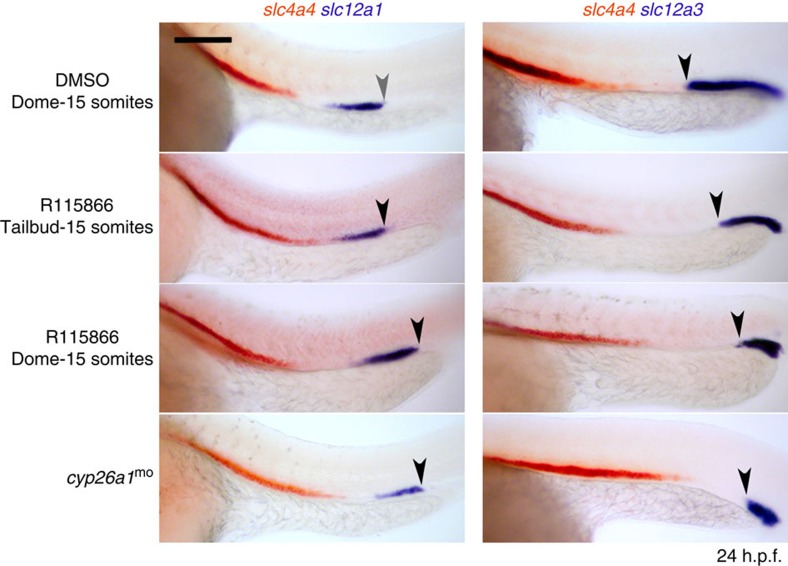
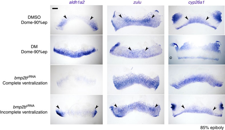
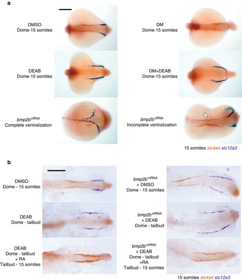
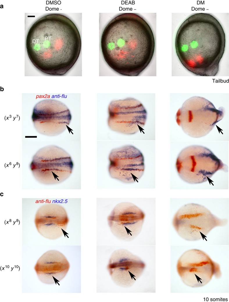
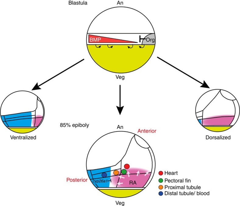
Despite the fundamental importance of patterning along the dorsal-ventral (DV) and anterior-posterior (AP) axes during embryogenesis, uncertainty exists in the orientation of these axes for the mesoderm. Here we examine the origin and formation of the zebrafish kidney, a ventrolateral mesoderm derivative, and show that AP patterning of the non-axial mesoderm occurs across the classic gastrula stage DV axis while DV patterning aligns along the animal-vegetal pole. We find that BMP signalling acts early to establish broad anterior and posterior territories in the non-axial mesoderm while retinoic acid (RA) functions later, but also across the classic DV axis. Our data support a model in which RA on the dorsal side of the embryo induces anterior kidney fates while posterior kidney progenitors are protected ventrally by the RA-catabolizing enzyme Cyp26a1. This work clarifies our understanding of vertebrate axis orientation and establishes a new paradigm for how the kidney and other mesodermal derivatives arise during embryogenesis.


Figures

Figure 1
Figure 1. Inhibition of RA signalling affects pronephros segmentation during gastrulation.
Whole-mount double in situ hybridization analysis at 24 hours post fertilisation (h.p.f.) for nephron segment markers slc4a4 (red), slc12a1 and slc12a3 (purple) treated with BMS189453, DEAB or DMSO (vehicle control). Embryos are shown as dorsal views with anterior to the left. White arrows indicate posterior end of slc4a4+ domain and black arrows indicate the boundary between the DE and DL segments. Scale bar, 100 μm.
Figure 2
Figure 2. Gene expression analysis of late gastrula embryos reveals distinct ventroposterior and dorsoanterior territories.
(a) Whole-mount in situ hybridization analysis for various mesodermal markers as indicated. Embryos were flat-mounted and are orientated with the animal side at the top and vegetal side at the bottom. Only mesodermal expression is highlighted with the exception of bmp4, which is expressed in the epiblast (expression in the hypoblast is unknown). (b) Schematic representation of the late gastrula mesodermal subdivisions. ALM, anterior lateral mesoderm; PPM, posterior paraxial mesoderm. Scale bar, 200 μm.
Figure 3
Figure 3. The zulu marker is highly expressed in the PLM.
(a) Whole-mount stage series in situ hybridization detecting gene expression of zulu suggest the zuluhigh PLM region at 85% epiboly forms the zuluhigh PLM at somitogenesis stages. All embryos are flat-mounted, 85% epiboly embryos are orientated with animal pole on top and vegetal pole at the bottom, later stages are dorsal views with anterior to the left. (b) Whole-mount double in situ hybridization at the 10-somites stage to detect transcripts for zulu and pax2a/gata1/nkx2.5/tbx5a. All embryos are flat-mounted and shown in dorsal views with anterior to the left. Scale bars, 200 μm.
Figure 4
Figure 4. Fate-mapping analysis of late gastrula embryos show proximal tubule and pectoral fin progenitors are juxtaposed to the aldh1a2high domain.
(a) Cells on the left-hand side of embryos at the 85% epiboly stage were lineage-labelled at various co-ordinates on the Cartesian grid shown (embryos are orientated with animal pole towards the top and dorsal side to the right). At the 10-somites stage, co-localization of uncaged cells and kidney (pax2a+), blood (gata1+), pectoral fin (tbx5a+) and heart (nkx2.5+) tissues was determined by whole-mount double in situ hybridization. Labelled cells on the right-hand side of the embryo (white asterisk) are the result of the laser passing through the embryo and uncaging cells on the contralateral side. Embryos are flat-mounted with anterior to the left (note: tbx5a is stained purple due to being a weaker probe). (b) Top schematic shows a flat-mounted embryo with the highlighted region corresponding to the panels below and the positions that label progenitors of the proximal (PT) and distal (DT) tubules, pectoral fin (PF) and heart (H). Bottom panels show whole-mount double in situ hybridization of these uncaged populations (red) relative to the expression domains of aldh1a2, cyp26a1 and zulu (purple) at 85% epiboly. (c) Lateral views of 70% (left) and 80% (centre) epiboly-stage embryos double stained for aldh1a2 (purple/black) and pax2a (red) transcripts. Higher magnified view of the indicated region is shown in the right-hand panel. A, animal pole; V, vegetal pole. Schematic at the bottom represents an outline of PLM. IM, intermediate mesoderm; LPM, lateral plate mesoderm. Scale bar in a, 100 μm in top panels and 200 μm in lower panels. Scale bars in b and c, 100 μm.
Figure 5
Figure 5. Knock down of Cyp26a1 anteriorizes the kidney.
Whole-mount double in situ hybridization analysis at 24 hours post fertilisation (h.p.f.) for nephron segment markers slc4a4 (orange), slc12a1 and slc12a3 (purple) in embryos injected with cyp26a1 morpholino or treated with R115866 or DMSO (vehicle control). Embryos are shown as lateral views with anterior to the left. Arrowhead indicates junction between the DE and DL segments. Scale bar, 100 μm.
Figure 6
Figure 6. BMP signalling regulates the size of aldh1a2 and cyp26a1 and zulu expression domains.
Whole-mount in situ hybridization analysis for aldh1a2, cyp26a1 and zulu at the 85% epiboly stage in embryos treated with DM dihydrochloride, DMSO (vehicle control) or injected with bmp2b mRNA. Embryos are shown flat-mounted and orientated with the animal pole at the top and the vegetal pole at the bottom. Scale bar, 200 μm.
Figure 7
Figure 7. RA acts downstream of the BMP signalling pathway during kidney patterning.
(a) Whole-mount double in situ hybridization analysis for nephron segment markers slc4a4 (red) and slc12a3 (purple) in embryos at the 15-somites stage injected with bmp2b mRNA or treated with DM dihydrochloride, DEAB and DMSO (vehicle control) at the indicated stages. Embryos are shown as dorsal views with anterior to the left. (b) Embryos were treated as shown and stopped at the 15-somites stage for double in situ hybridization staining for the anterior pronephros marker slc4a4 and the distal pronephros marker slc12a3. All panels are dorsal views of flatmounted embryos. Scale bar, 200 μm.
Figure 8
Figure 8. Effects of BMP and RA on cell fate and cell movement.
(a) Embryos were lineage labelled for distal (DT) and proximal (PT) cells (green) at 85% epiboly. One hour later the positions of these cells were analysed (pseudo-coloured red) and overlayed with the initial lineage labelling. Embryos are lateral views with anterior towards the top. (b) Uncaging cells at (x3, y7) or (x6, y8) at 85% epiboly labelled DT and PT regions, respectively, of the pax2a+ intermediate mesoderm in DMSO- and DEAB-treated 10-somites stage embryos. In DM-treated embryos, (x3, y7) uncaging labelled anterior intermediate mesoderm and a region we predict corresponds to anterior paraxial mesoderm. DM-treated embryos uncaged at (x6, y8) labelled a region of the embryo that likely was composed solely of anterior paraxial mesoderm. Embryos are shown as dorsal views with anterior to the left. (c) Uncaging at (x8, y8) labelled cells just posterior to heart progenitors in DMSO control embryos at the 10-somites stage, but labelled within the posterior domain of expanded heart progenitors (nkx2.5+ cells) in DEAB-treated embryos. Similarly, (x10, y10) labelled heart progenitors in 10-somites stage DMSO control embryos, but mainly labelled the anterior region of the expanded nkx2.5+ domain in DEAB-treated embryos. In DM-treated embryos, heart progenitors were found to sparsely populate the ventral side of the embryo, and (x8, y8) and (x10, y10) labelling at 85% epiboly gave rise to progressively more anterior structures that were positioned close to the dorsal midline. Embryos are shown as dorsal views slightly oblique such that the anterior head region is in view. Scale bars, 100 μm. Arrows indicate uncaged tracer.
Figure 9
Figure 9. Model showing how a radial field of RA is proposed to regulate AP patterning of the mesoderm downstream of BMP signalling.
Top schematic depicts a pregastrula embryo showing the equatorial mesendoderm, the organizer/shield region, and the proposed BMP activity gradient. Small curved arrows indicate the involution movements of the mesoderm at the margin. Bottom schematic depicts a late gastrula-stage embryo showing axial (prechordal plate and notochord), anterior and posterior mesodermal domains (see text for details), the positions of fate-mapped progenitors and the proposed radial field of RA relative to cyp26a1 expression. An, animal; Veg, vegetal.

Similar articles

Cited by

References

    1. Lane M. C. & Sheets M. D. Rethinking axial patterning in amphibians. Dev. Dyn. 225, 434–447 (2002). - PubMed
    1. Kumano G. & Smith W. C. Revisions to the Xenopus gastrula fate map: implications for mesoderm induction and patterning. Dev. Dyn. 225, 409–421 (2002). - PubMed
    1. Warga R. M. & Nusslein-Volhard C. Origin and development of the zebrafish endoderm. Development 126, 827–838 (1999). - PubMed
    1. Szeto D. P. & Kimelman D. The regulation of mesodermal progenitor cell commitment to somitogenesis subdivides the zebrafish body musculature into distinct domains. Genes Dev. 20, 1923–1932 (2006). - PMC - PubMed
    1. Fauny J. D., Thisse B. & Thisse C. The entire zebrafish blastula-gastrula margin acts as an organizer dependent on the ratio of Nodal to BMP activity. Development 136, 3811–3819 (2009). - PubMed

Publication types

MeSH terms

LinkOut - more resources