. 2016 Oct;15(5):914-23.
doi: 10.1111/acel.12500. Epub 2016 Jul 12.

Beta-amyloid 1-42 monomers, but not oligomers, produce PHF-like conformation of Tau protein

Affiliations

Beta-amyloid 1-42 monomers, but not oligomers, produce PHF-like conformation of Tau protein

Giusi Manassero et al. Aging Cell. 2016 Oct.

Abstract

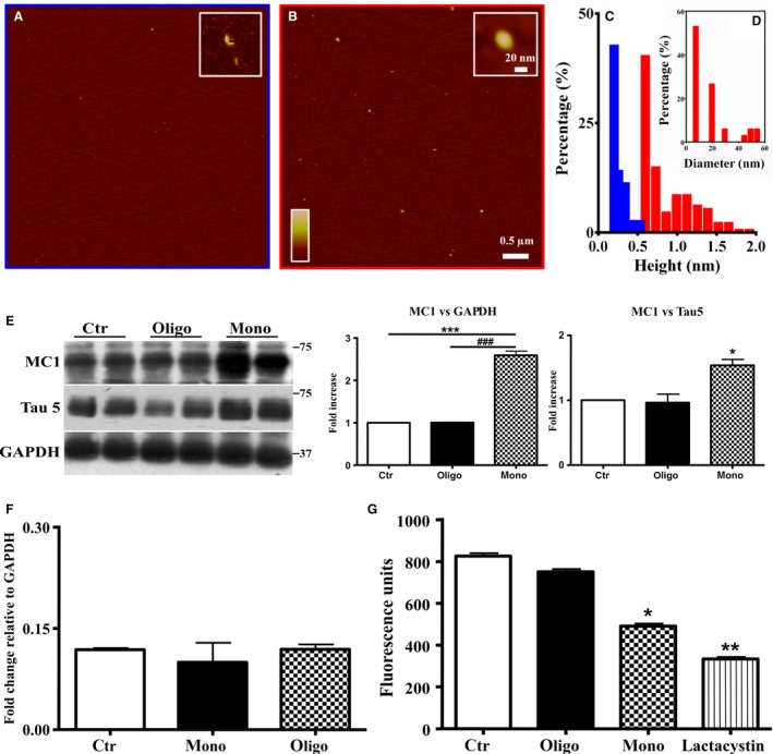
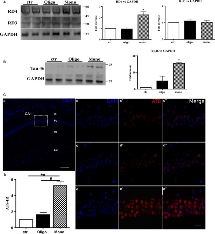
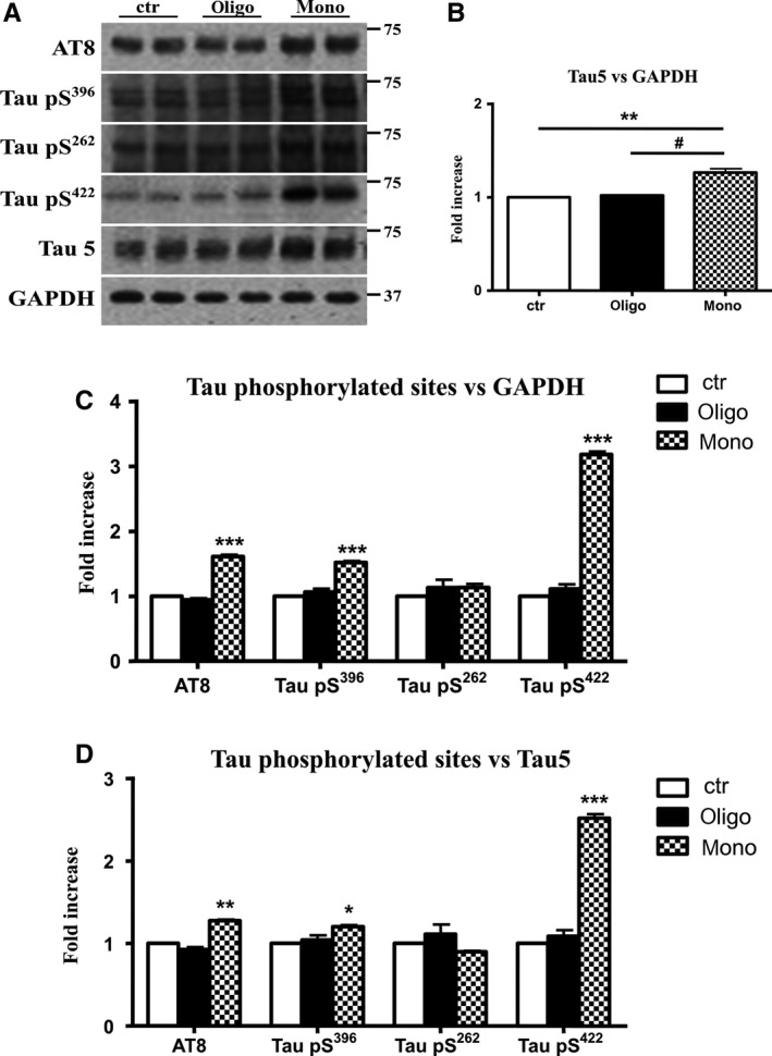
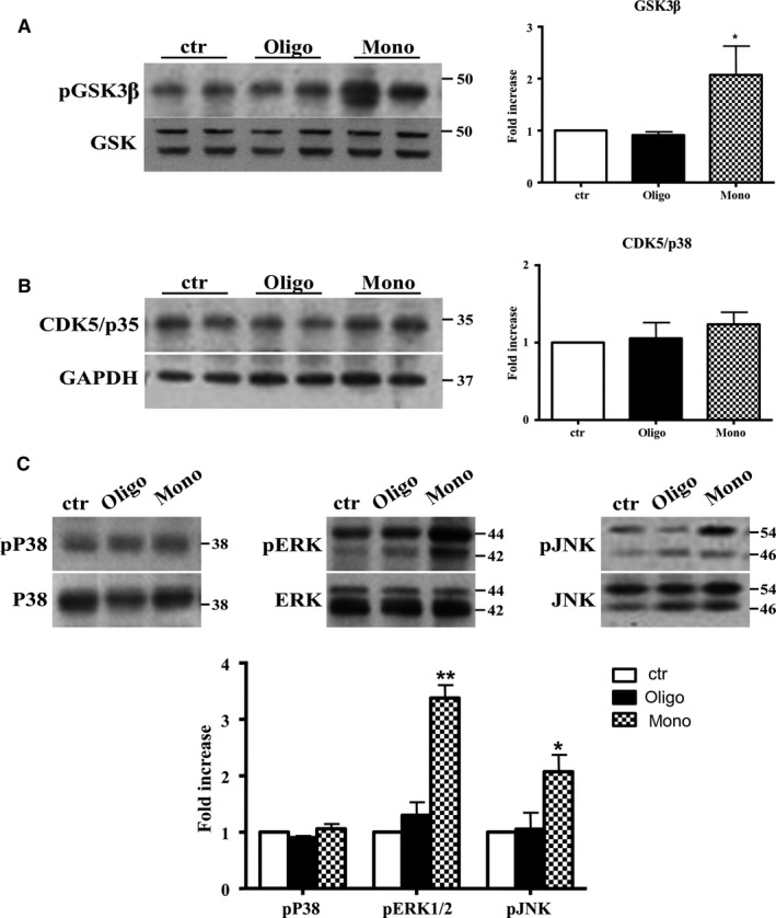
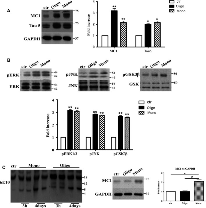
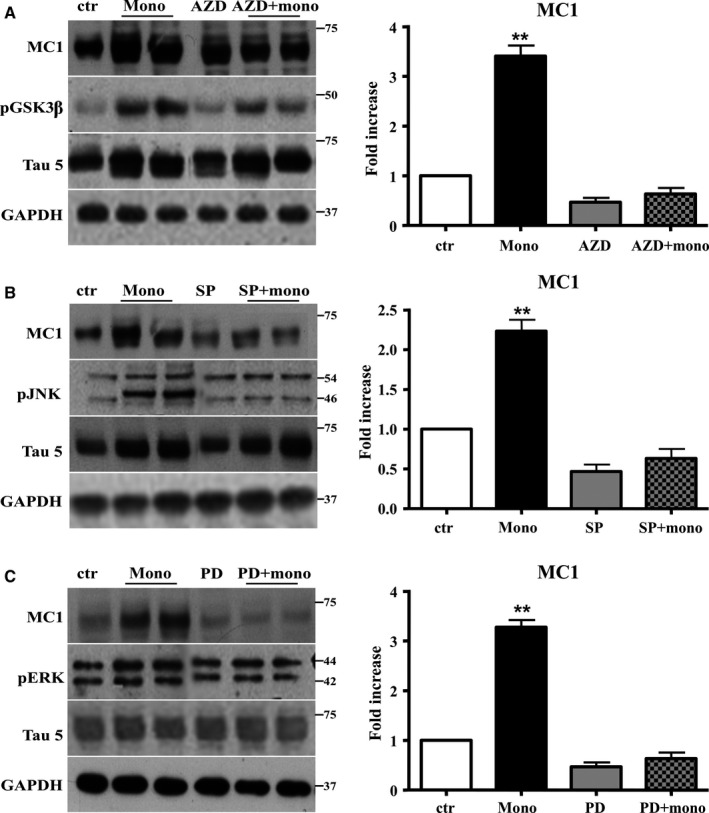
The mechanistic relationship between amyloid β1-42 (Aβ1-42) and the alteration of Tau protein are debated. We investigated the effect of Aβ1-42 monomers and oligomers on Tau, using mice expressing wild-type human Tau that do not spontaneously develop Tau pathology. After intraventricular injection of Aβ1-42, mice were sacrificed after 3 h or 4 days. The short-lasting treatment with Aβ monomers, but not oligomers, showed a conformational PHF-like change of Tau, together with hyperphosphorylation. The same treatment induced increase in concentration of GSK3 and MAP kinases. The inhibition of the kinases rescued the Tau changes. Aβ monomers increased the levels of total Tau, through the inhibition of proteasomal degradation. Aβ oligomers reproduced all the aforementioned alterations only after 4 days of treatment. It is known that Aβ1-42 monomers foster synaptic activity. Our results suggest that Aβ monomers physiologically favor Tau activity and dendritic sprouting, whereas their excess causes Tau pathology. Moreover, our study indicates that anti-Aβ therapies should be targeted to Aβ1-42 monomers too.

Keywords: Alzheimer's disease; MAPK; PHF; beta-amyloid; hTau mice; tau protein.


Figures

Figure 1
Figure 1
Aβ1‐42 monomers induce a conformational change of Tau protein. (A‐B) Analysis of the molecular assemblies of monomeric and oligomeric forms of Aβ 1‐42. (C) Representative atomic force microscopy (AFM) images of monomeric (initial state) and oligomeric assemblies, respectively. (D) Histogram representation of the heights of monomeric (blue bars) and oligomeric (red bars) assemblies, respectively. Distribution of the diameter histogram of oligomeric assemblies. (E) Representative Western blot of brain extracts from control (saline) and treated (Aβ1‐42 peptides by ICV for 3 h) mice (2‐month‐old) using a conformational Tau antibody (MC1) and a total Tau antibody (Tau5) for detection. An antibody raised against GAPDH or Tau 5 served as loading control. Densitometric quantification shows an increase of the total protein level of both MC1 and Tau5 induced by monomers. (F) Quantitative real‐time PCR (qPCR) analysis on total RNA extracted from brains of control and treated mice. Both treatments were not able to affect gene transcription. GAPDH mRNA expression was used as the internal standard. (G) Proteasomal activity assay on brain extracts from control and treated mice. Monomers halved the proteasomal activity. Lactacystin was used as a positive control. (H) Representative Western blot of brain extracts from control (saline) and treated with Aβ1‐42 monomeric as well as oligomeric depsipeptides by ICV for 3 h) mice (2‐month‐old) using a conformational Tau antibody (MC1). An antibody raised against GAPDH served as loading control. Densitometric quantification shows an increase of the total protein level of both MC1 induced by depsi‐monomers. The data are mean ± standard error of the mean (SEM), *P < 0.05; **P < 0.01 vs. control by one‐way ANOVA followed by Bonferroni post hoc test, n = 6.
Figure 2
Figure 2
Aβ1‐42 monomers produce alternative splicing, insoluble Tau aggregates, and hyperphosphorylation of Tau protein. (A) Representative Western blot of brain extracts from control (saline) and treated (Aβ1‐42 peptides by ICV for 3 h) mice (2‐month‐old) using a 4‐repeat Tau isoform RD4 and 3‐repeat Tau isoform RD3 for detection. An antibody raised against GAPDH served as loading control. Densitometric quantification shows an increase of the isoform with four binding domains mediated by monomers. (B) Representative Western blot of insoluble Tau fraction by sarkosyl detergent technique extracts from control (saline) and treated (Aβ1‐42 peptides by ICV for 3 h) mice (2‐month‐old) using a Tau 46 antibody for detection. An antibody raised against GAPDH served as loading control. Only after treatment with monomeric preparations was present a band at approximately 75 kDa molecular weight. (C) a. Schematic image of the mouse hippocampus CA1 region (coronal section). b. Quantification of AT8‐immunoreactive (IR) cells by imagej NIH software. The data are mean ± standard error of the mean (SEM),**P < 0.01 vs. ctr and #P < 0.05 vs. oligo by one‐way ANOVA followed by Bonferroni post hoc test; n = 9. c–e″. Higher magnifications of the box area in a. showing pyramidal layer of control and treated mice (Aβ1‐42 peptides by ICV for 3 h) stained with an antibody that recognizes PHF‐Tau (AT8 in red). Nuclei are counterstained with DAPI (blue). Scale bars: 100 μm in a, 20 μm in c–e″.
Figure 3
Figure 3
Aβ1‐42 monomers promote phosphorylation at particular sites that have been related to Alzheimer's disease (AD) progression. Representative Western blot of brain extracts (20 μg protein) from control (saline) and treated (Aβ1‐42 peptides by ICV for 3 h) mice using antibodies specific for the detection four pathological Tau phosphorylation sites: AT8, pS396, pS262, and pS422. An antibody raised against GAPDH or Tau 5 served as loading control. Densitometric quantification shows an increase of the total protein level of AT8, pS396, and pS422 induced by monomers while monomeric and oligomeric preparations did not change pS262 expression. The data are mean ± standard error of the mean (SEM), *P < 0.05; ***P < 0.001 vs. control by one‐way ANOVA followed by Bonferroni post hoc test, n = 6.
Figure 4
Figure 4
Aβ1‐42 monomers affect Tau phosphorylation through GSK3β, ERK1/2, and JNK kinases activation. Representative Western blot of brain extracts from control (saline) and treated mice using pGSK3β (A), CDK5/p35 (B), pP38, pERK1/2, and pJNK (C) antibodies for detection. Densitometric quantification shows an increase in the total protein level of pGSK3β induced by monomers (A), while CDK5/p35 was not modified by any treatments (B). (A,B) An antibody raised against GAPDH was used as loading control. The data are mean ± standard error of the mean (SEM), *P < 0.05 vs. control by one‐way ANOVA followed by Bonferroni post hoc test, n = 6 for each kinase. (C) pP38, pERK1/2, and pJNK levels were standardized against their respective total protein amount. Densitometric quantification shows an increase of ERK1/2 and JNK activity due to monomers, while p38 was not involved by any treatments. The data are mean ± standard error of the mean (SEM), *< 0.05; **< 0.01 vs. control by two‐way ANOVA followed by Bonferroni post hoc test, n = 3 for each kinase.
Figure 5
Figure 5
After 4 days of treatment, Aβ1‐42 oligomers and the monomers show a similar effect on Tau protein and phospho‐kinases. Representative Western blot of brain extracts from control (saline) and treated mice using MC1 and Tau5 (A), pERK1/2, pJNK, and pGSK3β (B) antibodies for detection. (A) Densitometric quantification shows an increase in the total protein level of both MC1 and Tau5 induced by both preparations. An antibody raised against GAPDH served as loading control. The data are mean ± standard error of the mean (SEM), *< 0.05, **< 0.01 vs. control by one‐way ANOVA followed by Bonferroni post hoc test, n = 3. (B) Densitometric quantification shows an increase of ERK1/2 and JNK activity due to both monomers and oligomers treatments. pERK1/2 and pJNK levels were standardized against their respective total protein amount. The data are mean ± SEM, **< 0.01 vs. control by two‐way ANOVA followed by Bonferroni post hoc test, n = 3 for each kinase. (C) Representative Western blot of the aggregation state of Aβ1‐42 in brain tissue of hTau mice injected after 3 h or 4 days with both preparations using the 6E10 antibody. As shown, the aggregation state of oligomeric preparation was different in the short and in the longer treatment: After 3 h, a band of approximately 12 Kda was observed, whereas after 4 days, this band was no more detectable. The data are mean ± SEM, **< 0.01 vs. control by two‐way ANOVA followed by Bonferroni post hoc test, n = 3.
Figure 6
Figure 6
The activation of JNK, ERK1/2, and GSK is required to mediate the conformational change of Tau protein induced by Aβ1‐42 monomers. Representative Western blot of brain extracts from control and pretreated or not with the GSK3β inhibitor AZD1080, ERK inhibitor PD98059, and JNK inhibitor SP600125 before 3 h injection with Aβ1‐42 Tau mice using MC1 (A‐C), pGSK3β (A), pJNK (B), and pERK1/2 (C) antibodies for detection. An antibody raised against GAPDH or Tau5 served as loading control. Densitometric quantification shows that each pretreatment was followed by a complete inhibition of the pathway and by the complete reversion of Tau conformational change (A–C). The data are mean ± standard error of the mean (SEM), **< 0.01 vs. control by one‐way ANOVA followed by Bonferroni post hoc test, n = 6 for each kinase.

References

    1. Alonso A, Zaidi T, Novak M, Grundke‐Iqbal I, Iqbal K (2001) Hyperphosphorylation induces self‐assembly of tau into tangles of paired helical filaments/straight filaments. Proc. Natl Acad. Sci. USA 98, 6923–6928. - PMC - PubMed
    1. Ando K, Maruko‐Otake A, Ohtake Y, Hayashishita M, Sekiya M, Iijima KM (2016) Stabilization of microtubule‐unbound tau via tau phosphorylation at Ser262/356 by Par‐1/MARK contributes to augmentation of ad‐related phosphorylation and Aβ42‐induced tau toxicity. PLoS Genet. 12, e1005917. - PMC - PubMed
    1. Andorfer C, Kress Y, Espinoza M, de Silva R, Tucker KL, Barde YA, Duff K, Davies P (2003) Hyperphosphorylation and aggregation of tau in mice expressing normal human tau isoforms. J. Neurochem. 86, 582–590. - PubMed
    1. Beeg M, Stravalaci M, Bastone A, Salmona M, Gobbi M (2011) A modified protocol to prepare seed‐free starting solutions of amyloid‐β (Aβ)₁₋₄₀ and Aβ₁₋₄₂ from the corresponding depsipeptides. Anal. Biochem. 411, 297–299. - PubMed
    1. Bero AW, Yan P, Roh JH, Cirrito JR, Stewart FR, Raichle ME, Lee JM, Holtzman DM (2011) Neuronal activity regulates the regional vulnerability to amyloid‐β deposition. Nat. Neurosci. 14, 750–756. - PMC - PubMed

Publication types