. 2017 Oct;11(10):2912-2921.
doi: 10.1002/term.2194. Epub 2016 Jul 13.

Asymmetrical seeding of MSCs into fibrin-poly(ester-urethane) scaffolds and its effect on mechanically induced chondrogenesis

Affiliations

Asymmetrical seeding of MSCs into fibrin-poly(ester-urethane) scaffolds and its effect on mechanically induced chondrogenesis

Oliver F W Gardner et al. J Tissue Eng Regen Med. 2017 Oct.

Abstract

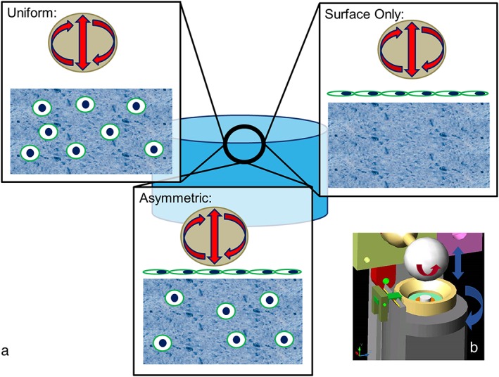
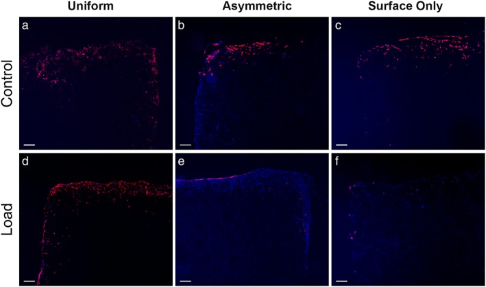
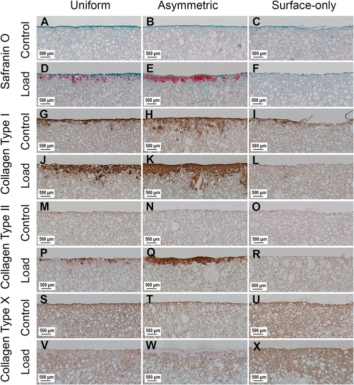
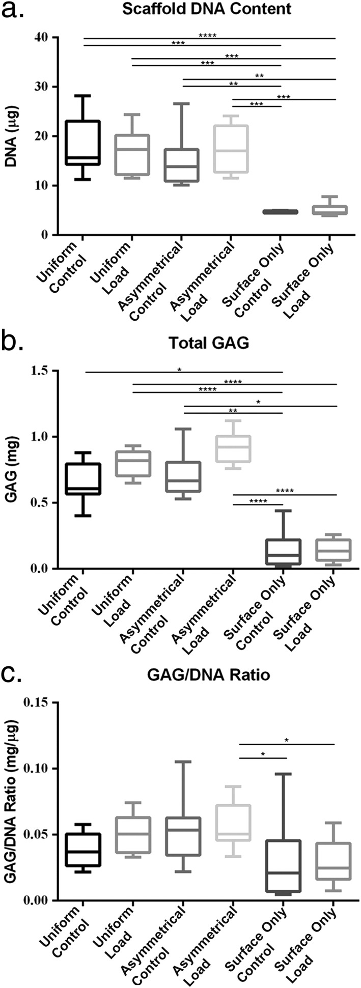
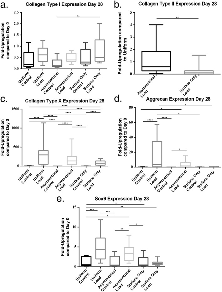
Mesenchymal stem cells (MSCs) are currently being investigated as candidate cells for regenerative medicine approaches for the repair of damaged articular cartilage. For these cells to be used clinically, it is important to understand how they will react to the complex loading environment of a joint in vivo. In addition to investigating alternative cell sources, it is also important for the structure of tissue-engineered constructs and the organization of cells within them to be developed and, if possible, improved. A custom built bioreactor was used to expose human MSCs to a combination of shear and compression loading. The MSCs were either evenly distributed throughout fibrin-poly(ester-urethane) scaffolds or asymmetrically seeded with a small proportion seeded on the surface of the scaffold. The effect of cell distribution on the production and deposition of cartilage-like matrix in response to mechanical load mimicking in vivo joint loading was then investigated. The results show that asymmetrically seeding the scaffold led to markedly improved tissue development based on histologically detectable matrix deposition. Consideration of cell location, therefore, is an important aspect in the development of regenerative medicine approaches for cartilage repair. This is particularly relevant when considering the natural biomechanical environment of the joint in vivo and patient rehabilitation protocols. Copyright © 2016 John Wiley & Sons, Ltd.

Keywords: bioreactor; cartilage repair; mesenchymal stem cell; multi-axial load; poly-ester-urethane; shear.


Conflict of interest statement

The authors have declared that there is no conflict of interest.

Figures

Figure 1
Figure 1
(a) Schematic showing the different seeding patterns used during this work. In Uniform 4 million cells were evenly seeded throughout scaffolds. In Asymmetrically seeded scaffolds, 3.6 million cells were seeded evenly throughout the scaffold and 400 000 cells seeded on the loaded surface of the scaffold. Surface Only scaffolds were not seeded with cells within the scaffold but only with 400 000 cells on the loaded surface. (b) Schematic showing the multi‐axial loading device used in the present study. The hip ball is lowered onto the white disc representing the scaffold. Compression is generated by raising and lowering the ball, and sliding friction is generated by rotating the ball, allowing for a sliding motion over the scaffold. [Colour figure can be viewed at http://wileyonlinelibrary.com]
Figure 2
Figure 2
Fluorescent images showing the location of labelled cells after 4 weeks of load. Images (a), (b) and (c) show control scaffolds from Uniform, Asymmetric and Surface Only seeded scaffolds respectively, while images (d), (e) and (f) show loaded scaffolds. Red fluorescence shows the membranes of labelled cells, blue shows nuclei counterstained with 4′,6‐diamidino‐2‐phenylindole (DAPI) (n = 1). Bar, 200 μm. [Colour figure can be viewed at http://wileyonlinelibrary.com]
Figure 3
Figure 3
Images showing the surface of scaffolds stained with Safranin O and immunohistochemical labelling for collagen type I, collagen type II and collagen type X after 4 weeks of culture. Images (a), (b) and (c) show Safranin O‐stained control scaffolds from Uniform, Asymmetric and Surface Only seeded scaffolds, respectively, while images (d), (e) and (f) show Safranin O‐stained loaded scaffolds. Images (g), (h) and (i) show control scaffolds labelled for collagen type I, and images (j) (k) and (l) show loaded scaffolds. Images (m), (n) and (o) show control scaffolds labelled for collagen type II and images (p), (q) and (r) show loaded scaffolds. Images (s), (t) and (u) show control scaffolds labelled for collagen type X and images (v), (w) and (x) show loaded scaffolds. [Colour figure can be viewed at http://wileyonlinelibrary.com]
Figure 4
Figure 4
Biochemical analysis of scaffolds after 4 weeks in culture. (a) Höchst 33528 dye was used to quantify the DNA in proteinase K digests of scaffolds. (b) Dimethylmethylene blue (DMMB) was used to determine the total amount of sulphated glycosaminoglycan (GAG) produced by mesenchymal stem cells (MSCs) from both the collected culture media and proteinase K scaffold digests. (c) The GAG/DNA ratio was calculated from total DNA and GAG values to show the production of GAG relative to the MSCs present in each group. Error bars represent standard deviation. Statistical significance was defined as P ≤ 0.05; *P ≤ 0.05, **P ≤ 0.001 and ***P ≤ 0.0001
Figure 5
Figure 5
Gene expression measured at day 28 of culture by real‐time polymerase chain reaction (PCR). (a) Collagen type I, (b) collagen type II, (c) collagen type X, (d) aggrecan and (e) Sox9. Error bars represent standard deviation. Statistical significance was defined as P ≤ 0.05; *P ≤ 0.05, **P ≤ P≤0.001 and ***P ≤ 0.0001

References

    1. Atala A, Bauer SB, Soker S et al 2006; Tissue‐engineered autologous bladders for patients needing cystoplasty. Lancet 367: 1241–1246. - PubMed
    1. Benya PD, Shaffer JD. 1982; Dedifferentiated chondrocytes reexpress the differentiated collagen phenotype when cultured in agarose gels. Cell 30: 215–224. - PubMed
    1. Boissard I, Bourban PE, Tami AE et al 2009; Nanohydroxyapatite/poly(ester urethane) scaffold for bone tissue engineering. Acta Biomater 5: 3316–3327. - PubMed
    1. Brittberg M, Lindahl A, Nilsson A et al 1994; Treatment of deep cartilage defects in the knee with autologous chondrocyte transplantation. N Engl J Med 331: 889–895. - PubMed
    1. Cherubino P, Grassi FA, Bulgheroni P, et al 2003; Autologous chondrocyte implantation using a bilayer collagen membrane: a preliminary report. J Orthop Surg (Hong Kong) 11: 10–15. - PubMed

Publication types

MeSH terms

LinkOut - more resources