. 2016 Aug;22(8):781-91.
doi: 10.1089/ten.TEC.2016.0066.

Development of a Full-Thickness Human Gingiva Equivalent Constructed from Immortalized Keratinocytes and Fibroblasts

Affiliations

Development of a Full-Thickness Human Gingiva Equivalent Constructed from Immortalized Keratinocytes and Fibroblasts

Jeroen K Buskermolen et al. Tissue Eng Part C Methods. 2016 Aug.

Abstract

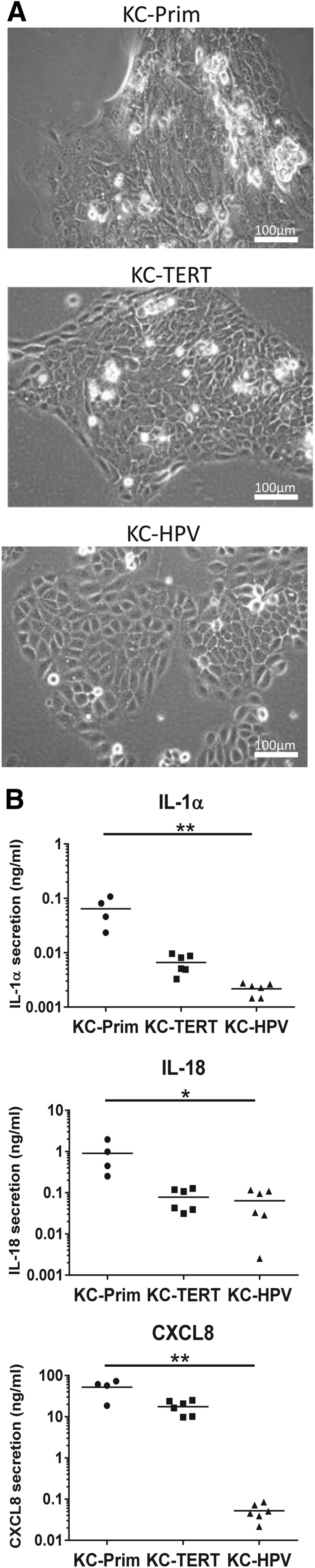
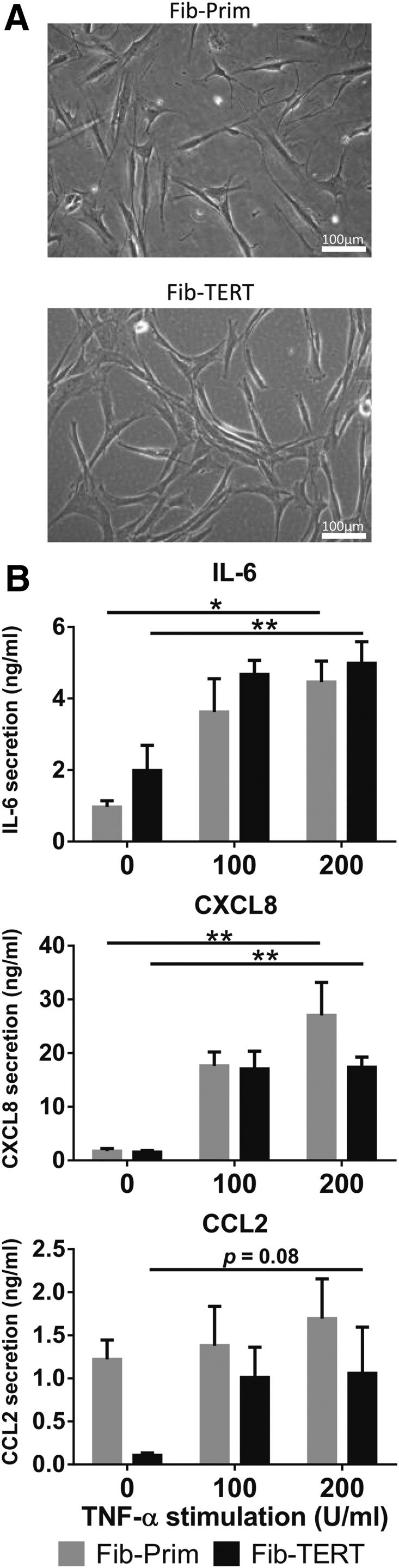
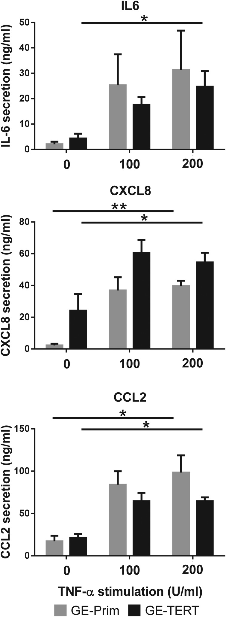
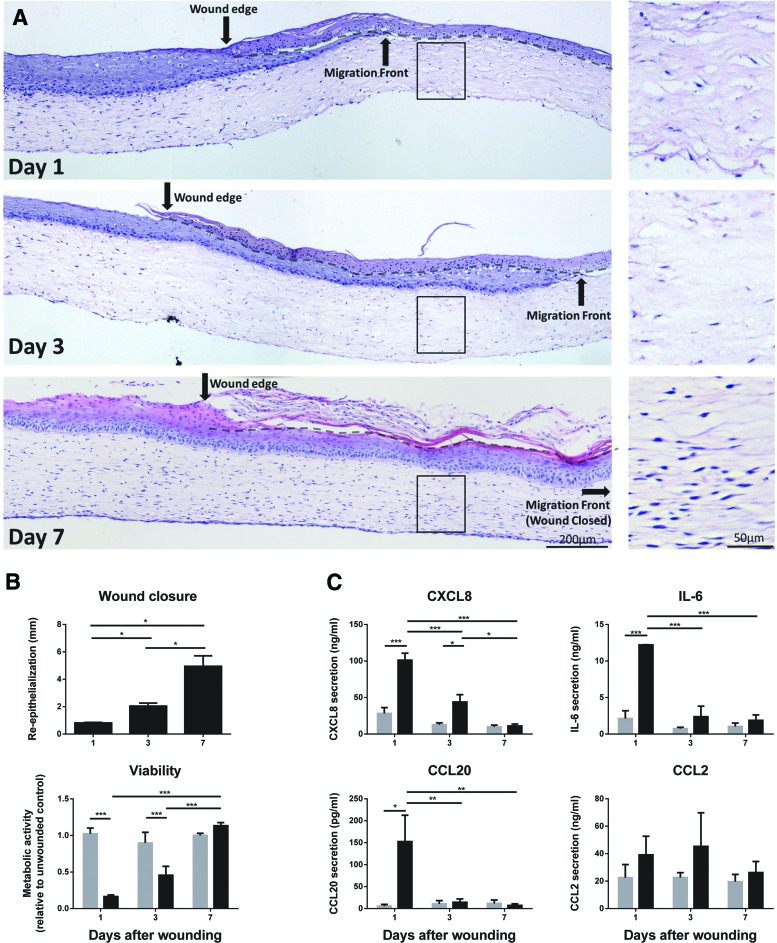
Organotypic models make it possible to investigate the unique properties of oral mucosa in vitro. For gingiva, the use of human primary keratinocytes (KC) and fibroblasts (Fib) is limited due to the availability and size of donor biopsies. The use of physiologically relevant immortalized cell lines would solve these problems. The aim of this study was to develop fully differentiated human gingiva equivalents (GE) constructed entirely from cell lines, to compare them with the primary cell counterpart (Prim), and to test relevance in an in vitro wound healing assay. Reconstructed gingiva epithelium on a gingiva fibroblast-populated collagen hydrogel was constructed from cell lines (keratinocytes: TERT or HPV immortalized; fibroblasts: TERT immortalized) and compared to GE-Prim and native gingiva. GE were characterized by immunohistochemical staining for proliferation (Ki67), epithelial differentiation (K10, K13), and basement membrane (collagen type IV and laminin 5). To test functionality of GE-TERT, full-thickness wounds were introduced. Reepithelialization, fibroblast repopulation of hydrogel, metabolic activity (MTT assay), and (pro-)inflammatory cytokine release (enzyme-linked immunosorbent assay) were assessed during wound closure over 7 days. Significant differences in basal KC cytokine secretion (IL-1α, IL-18, and CXCL8) were only observed between KC-Prim and KC-HPV. When Fib-Prim and Fib-TERT were stimulated with TNF-α, no differences were observed regarding cytokine secretion (IL-6, CXCL8, and CCL2). GE-TERT histology, keratin, and basement membrane protein expression very closely represented native gingiva and GE-Prim. In contrast, the epithelium of GE made with HPV-immortalized KC was disorganized, showing suprabasal proliferating cells, limited keratinocyte differentiation, and the absence of basement membrane proteins. When a wound was introduced into the more physiologically relevant GE-TERT model, an immediate inflammatory response (IL-6, CCL2, and CXCL8) was observed followed by complete reepithelialization. Seven days after wounding, tissue integrity, metabolic activity, and cytokine levels had returned to the prewounded state. In conclusion, immortalized human gingiva KC and fibroblasts can be used to make physiologically relevant GE, which resemble either the healthy gingiva or a neoplastic disease model. These organotypic models will provide valuable tools to investigate oral mucosa biology and can also be used as an animal alternative for drug targeting, vaccination studies, microbial biofilm studies, and testing new therapeutics.


Figures

<b>FIG. 1.</b>
FIG. 1.
Morphology and cytokine secretion of TERT-immortalized gingiva keratinocytes are similar to primary gingiva keratinocytes. Primary and immortalized human gingiva keratinocytes grown under conventional submerged culture conditions. (A) Phase contrast images of primary (KC-Prim), TERT-immortalized (KC-TERT), and HPV-immortalized (KC-HPV) human gingiva keratinocytes. (B) Secretion of IL-1α, IL-18, and CXCL8 by gingiva keratinocytes into culture supernatants over a 24-h culture period. Results are from four individual donors for KC-Prim and six different passages between passage 30 and 50 for the immortalized gingiva keratinocytes KC-TERT and KC-HPV; *p < 0.05; **p < 0.01; Kruskal–Wallis test.
<b>FIG. 2.</b>
FIG. 2.
Morphology and cytokine secretion of TERT-immortalized gingiva fibroblasts are similar to primary gingiva fibroblasts. Primary (Fib-Prim) and TERT-immortalized (Fib-TERT) human gingiva fibroblasts grown under conventional submerged culture conditions. (A) Phase contrast images of Fib-Prim and Fib-TERT. (B) Secretion of IL-6, CXCL8, and CCL2 into culture supernatants by Fib-Prim (gray bars) and Fib-TERT (black bars) exposed to rhTNF-α over 24 h. Data represent the average of at least three individual donors for Fib-Prim and four different passages of Fib-TERT between passage 20 and 30 ± SEM; *p < 0.05; **p < 0.01; two-way ANOVA for analysis of response to rhTNF-α exposure and Sidak's multiple comparison between Fib-Prim and Fib-TERT. SEM, standard error of mean.
<b>FIG. 3.</b>
FIG. 3.
Full-thickness gingiva equivalents (GE) constructed entirely from cell lines closely represent the native gingiva tissue architecture. GEs were constructed from primary human gingiva keratinocytes and fibroblasts (GE-Prim) or either TERT (GE-TERT)- or HPV (GE-HPV)-immortalized keratinocytes with TERT-immortalized fibroblasts as described in the Materials and Methods section. Figures are representative of at least three independent experiments. (A) Paraffin tissue sections (5 μm) were stained with hematoxylin and eosin (H&E) to visualize histology or immunohistochemically processed with antibodies directed against epithelial biomarkers (involucrin, K10, or K13), proliferation marker Ki-67, or the fibroblast biomarker vimentin (red immune staining). Scale bar represents 200 μm for the native gingiva biopsies and 100 μm for the GEs. (B) Immunohistochemistry of basement membrane proteins collagen type IV and laminin 5 on cryosections (5 μm). Scale bar represents 25 μm. Color images available online at www.liebertpub.com/tec
<b>FIG. 4.</b>
FIG. 4.
TERT-immortalized GE secrete inflammatory cytokines in response to rhTNF-α in a similar manner to GE constructed from primary cells. Primary (GE-Prim; gray bars) and TERT-immortalized (GE-TERT, black bars) human GEs were grown as described in the Materials and Methods section and then cultured for 24 h in the presence or absence of different concentrations of rhTNF-α. Cytokine IL-6, CXCL8, and CCL2 secretion into culture supernatants was determined with ELISA. Data represent the average of three individual donors for GE-Prim and three different passages of immortalized cells between passage 30 and 50 ± SEM; *p < 0.05; **p < 0.01; two-way ANOVA for analysis of response to rhTNF-α exposure and Sidak's multiple comparison between GE-Prim and GE-TERT. ELISA, enzyme-linked immunosorbent assay.
<b>FIG. 5.</b>
FIG. 5.
TERT-immortalized GE are functional in an in vitro wound-healing assay. Full-thickness freeze wounds were introduced into TERT-immortalized human GE and reepithelialization and fibroblast migration into the wound area as well as inflammatory cytokine secretion were assessed 1, 3, and 7 days after introducing the wound. (A) H&E staining of tissue sections derived from GE-TERT 1, 3, and 7 days after wounding. Black arrow indicates original wound edge; dotted line indicates the interface between the dead epithelial layer and the ingrowing epithelial layer. Inlay shows absence and repopulation of fibroblasts within the original wound in the collagen hydrogel. (B) Upper panel: Quantification of reepithelialization from the wound margins 1, 3, and 7 days after wounding. Lower panel; 3 mm diameter punch biopsies were obtained from the unwounded area (gray bar) and wounded area (black bar) of GE-TERT and analyzed with the MTT mitochondrial activity assay, which represents cell viability. (C) Cytokine secretion by unwounded (gray bars) and wounded (black bars) GE-TERT cultures. Culture supernatants were collected over a 24-h period between 0–1, 2–3, and 6–7 days after wounding and analyzed by ELISA. All figures represent the average of three independent experiments ± SEM; *p < 0.05; **p < 0.01; ***p < 0.001; two-way ANOVA for response between days and Sidak's multiple comparison between unwounded and wounded areas/cultures. Color images available online at www.liebertpub.com/tec

References

    1. Presland R.B., and Dale B.A. Epithelial structural proteins of the skin and oral cavity: function in health and disease. Crit Rev Oral Biol Med 11, 383, 2000 - PubMed
    1. Dongari-Bagtzoglou A., and Kashleva H. Development of a highly reproducible three-dimensional organotypic model of the oral mucosa. Nat Protoc 1, 2012, 2006 - PMC - PubMed
    1. Vriens A.P., et al. . Comparison of autologous full-thickness gingiva and skin substitutes for wound healing. Cell Transplant 17, 1199, 2008 - PubMed
    1. Kosten I.J., Buskermolen J.K., Spiekstra S.W., de Gruijl T.D., and Gibbs S. Gingiva equivalents secrete negligible amounts of key chemokines involved in langerhans cell migration compared to skin equivalents. J Immunol Res 2015, 627125, 2015 - PMC - PubMed
    1. Gibbs S., and Ponec M. Intrinsic regulation of differentiation markers in human epidermis, hard palate and buccal mucosa. Arch Oral Biol 45, 149, 2000 - PubMed

LinkOut - more resources