. 2016 Jul 12;7(4):e00915-16.
doi: 10.1128/mBio.00915-16.

Horizontal Transmission of Cytosolic Sup35 Prions by Extracellular Vesicles

Affiliations

Horizontal Transmission of Cytosolic Sup35 Prions by Extracellular Vesicles

Shu Liu et al. mBio. .

Abstract

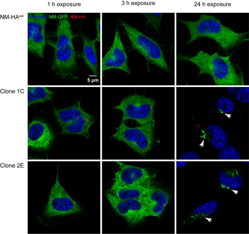
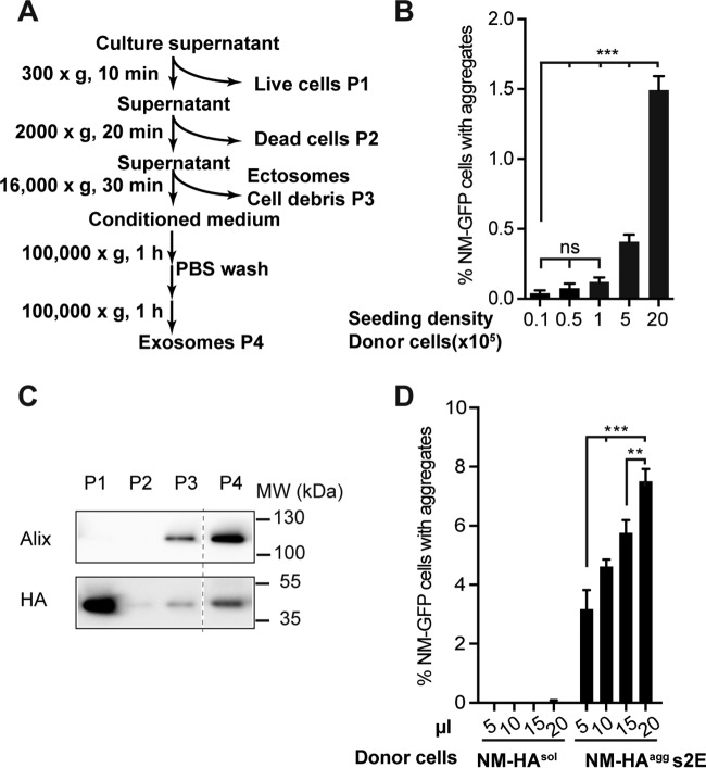
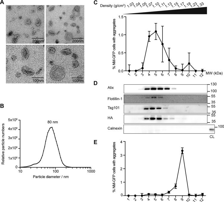
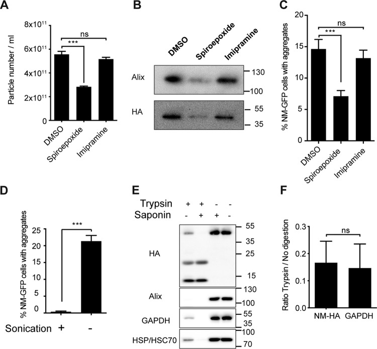
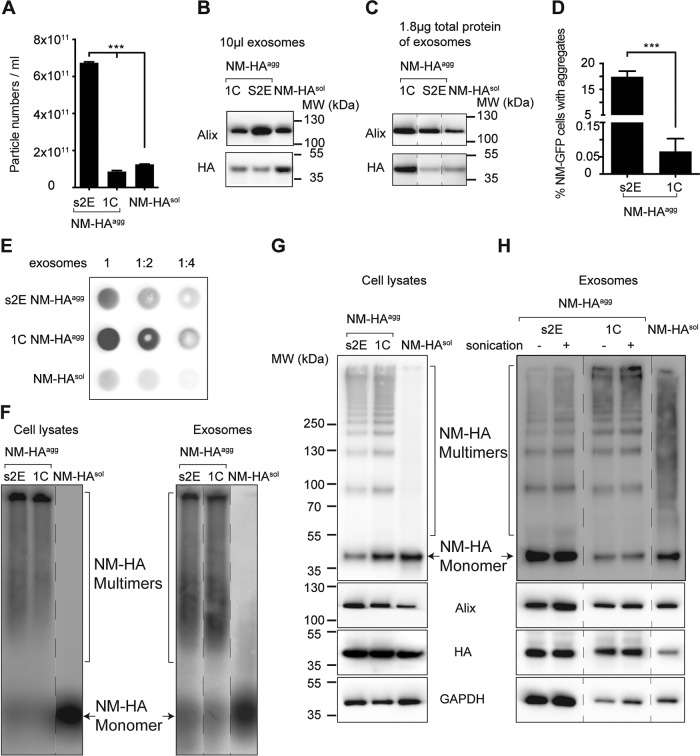
Prions are infectious protein particles that replicate by templating their aggregated state onto soluble protein of the same type. Originally identified as the causative agent of transmissible spongiform encephalopathies, prions in yeast (Saccharomyces cerevisiae) are epigenetic elements of inheritance that induce phenotypic changes of their host cells. The prototype yeast prion is the translation termination factor Sup35. Prions composed of Sup35 or its modular prion domain NM are heritable and are transmitted vertically to progeny or horizontally during mating. Interestingly, in mammalian cells, protein aggregates derived from yeast Sup35 NM behave as true infectious entities that employ dissemination strategies similar to those of mammalian prions. While transmission is most efficient when cells are in direct contact, we demonstrate here that cytosolic Sup35 NM prions are also released into the extracellular space in association with nanometer-sized membrane vesicles. Importantly, extracellular vesicles are biologically active and are taken up by recipient cells, where they induce self-sustained Sup35 NM protein aggregation. Thus, in mammalian cells, extracellular vesicles can serve as dissemination vehicles for protein-based epigenetic information transfer.

Importance: Prions are proteinaceous infectious particles that propagate by templating their quaternary structure onto nascent proteins of the same kind. Prions in yeast act as heritable epigenetic elements that can alter the phenotype when transmitted to daughter cells or during mating. Prion activity is conferred by so-called prion domains often enriched in glutamine and asparagine residues. Interestingly, many mammalian proteins also contain domains with compositional similarity to yeast prion domains. We have recently provided a proof-of-principle demonstration that a yeast prion domain also retains its prion activity in mammalian cells. We demonstrate here that cytosolic prions composed of a yeast prion domain are also packaged into extracellular vesicles that transmit the prion phenotype to bystander cells. Thus, proteins with prion-like domains can behave as proteinaceous information molecules that exploit the cellular vesicle trafficking machinery for intercellular long-distance dissemination.


Figures

FIG 1
FIG 1
Murine N2a cells harboring cytosolic Sup35 NM-HA aggregates secrete infectious NM prions. Shown are confocal images of recipient N2a NM-GFP cells (green) exposed to the 100,000 × g pelleted fractions derived from conditioned medium of NM-HA aggregate-producing cell clones 2E and 1C or control donor cells expressing soluble NM-HA. Recipient cells were fixed 1, 3, and 24 h posttreatment with pellet fractions. NM-GFP aggregate induction was observed after 24 h with pellet fractions derived from media of both clones but not from donor cells expressing NM-HAsol. NM-HA was stained with anti-HA antibody (red), and nuclei were counterstained with Hoechst (blue). NM-GFP aggregates are indicated by arrows. For images showing cells with NM-GFP aggregates, the microscopy setting had to be adjusted to prevent the overexposure of highly fluorescent aggregates. Note that no colocalization of internalized NM-HA seeds and NM-GFP aggregates was observed. Scale bar, 5 µm.
FIG 2
FIG 2
Infectious NM prions are associated with the exosomal fraction. (A) Differential centrifugation protocol to enrich for exosomes. Donor cells were grown in exosome-depleted medium. (B) Cell-based aggregate induction assay. Conditioned medium derived from NM-HAagg clone s2E plated at different densities induced NM-GFP aggregation in recipient cells in a dose-dependent manner. Shown is the mean ± SD (n = 3). Statistical analysis was performed by one-way ANOVA. ***, P < 0.001. (C) Western blot analysis of pellet fractions P1 to P4 isolated from conditioned medium of s2E cells according to the scheme in panel A. Alix served as a marker protein for exosomes. Anti-HA antibody was used to detect NM-HA. Additional lanes were excised for presentation purposes (dotted line). (D) Cell-based aggregate induction assay using the exosome-enriched P4 fraction isolated from the media of NM-HAsol and NM-HAagg s2E cells. The pellet was dissolved in PBS, and 5 to 20 µl was added to recipient NM-GFPsol cells. The number of recipient cells with induced aggregates was determined 16 h postexposure. The results shown are means ± SD (n = 4). **, P < 0.01; ***, P < 0.001; one-way ANOVA.
FIG 3
FIG 3
NM-HA and prion infectivity cofractionate with exosomes. (A) Transmission EM of the P4 fraction from donor clone s2E reveals the typical exosomal shape and dimension. Scale bars: top, 200 nm; bottom, 100 nm. (B) Size distribution of vesicles in the P4 fraction from media of donor clone s2E. (C) Fractionation of the exosome-enriched P4 fraction from medium of clone s2E by OptiPrep gradient centrifugation. Twelve fractions were collected and analyzed for aggregate-inducing activity in recipient NM-GFPsol cells (n = 3). (D) OptiPrep density gradient fractions used in panel C were subjected to Western blot analysis to test for the distribution of NM-HA and exosomal marker proteins Alix, Tsg101, and Flotillin-1. Calnexin served as a marker protein for the endoplasmic reticulum. s2E cell lysate (CL) was loaded as a control for calnexin detection. Note that none of the OptiPrep fractions contained calnexin, excluding organelle contamination. Immunoblotting with anti-HA antibodies revealed that NM-HA cofractionated with exosomal markers. (E) Fractionation of in vitro-formed NM fibrils by OptiPrep density gradient centrifugation. The different fractions were analyzed for aggregate-inducing activity in recipient NM-GFPsol cells. The highest induction rates were observed for fraction 9. The results shown are means ± SD (n = 3).
FIG 4
FIG 4
Exosomes serve as carriers of prion infectivity. (A) Determination of particle numbers. Donor clone s2E was treated with 5 µM spiroepoxide, 10 µM imipramine, or solvent control DMSO. Media were collected after 72 h, and exosome-enriched P4 fractions were isolated. The results shown are means ± SD (n = 3; ***, P < 0.001; ns, no significant difference; one-way ANOVA). (B) Samples were subjected to Western blot analysis to determine the levels of Alix and NM-HA. The values to the right are molecular sizes in kilodaltons. (C) Samples were analyzed for aggregate-inducing activity in recipient cells expressing NM-GFPsol. The results shown are means ± SD (n = 6; ***, P < 0.001; ns, no significant difference; one-way ANOVA). (D) Aliquots of the P4 fraction isolated from donor clone s2E were sonicated to disrupt exosomal membranes. Sonicated/nonsonicated samples were then subjected to the cell-based aggregate induction assay. The results shown are means ± SD (n = 6; ***, P < 0.0001; unpaired Student t test). (E) Western blot analysis of the P4 fraction (clone s2E) subjected to limited proteolysis in the presence or absence of saponin. GAPDH and Hsp70 served as markers of intraluminal proteins, and Alix is a protein associated with the exosomal membrane. (F) Fold difference in signal intensity of full-length NM-HA and GAPDH bands (with or without trypsin treatment) shown in panel E. The results shown are means ± SD (n = 6; ***, P < 0.001; ns, no significant difference; unpaired Student t test).
FIG 5
FIG 5
Aggregation state of NM-HA associated with exosomes. (A) Vesicle concentrations in the P4 fractions derived from media of NM-HAagg clones s2E and 1C and NM-HAsol cells measured by ZetaView nanoparticle tracking analysis. The results shown are means ± SD (n = 3; ***, P < 0.001; one-way ANOVA). (B, C) Western blot analyses of P4 fractions loaded at comparable volumes of isolated exosomes or adjusted to comparable total protein levels. The positions of the lanes were switched for presentation purposes (dashed lines). (D) Cell-based aggregate induction assay. Percentages of recipient cells with NM-GFPagg induced by P4 exosomal fractions of donor clones 1C and s2E are shown. The results shown are means ± SD (n = 6; ***, P < 0.0001; unpaired Student t test). (E) Filter trap assay using P4 fractions isolated from clones s2E and 1C and NM-HAsol control cells. Shown at the top are the dilutions used. (F) SDD-AGE analysis of cell lysates and exosome fractions derived from NM-HAsol cells and NM-HAagg clones s2E and 1C. (G, H) Glutaraldehyde cross-linking with cell lysates and exosomes from NM-HAsol cells and clones s2E and 1C (top). The same amount of sample without cross-linking was also loaded as a control and analyzed for Alix, HA, and GAPDH protein levels (bottom). Extra marker lanes were removed for presentation (dashed lines).

References

    1. Prusiner SB. 1982. Novel proteinaceous infectious particles cause scrapie. Science 216:136–144. doi: 10.1126/science.6801762. - DOI - PubMed
    1. Prusiner SB. 1998. Prions. Proc Natl Acad Sci U S A 95:13363–13383. doi: 10.1073/pnas.95.23.13363. - DOI - PMC - PubMed
    1. Moore RA, Vorberg I, Priola SA. 2005. Species barriers in prion diseases—brief review. Arch Virol Suppl 2005:187–202. - PubMed
    1. Heikenwalder M, Prinz M, Heppner FL, Aguzzi A. 2004. Current concepts and controversies in prion immunopathology. J Mol Neurosci 23:3–12. doi: 10.1385/JMN:23:1-2:003. - DOI - PubMed
    1. Vella LJ, Sharples RA, Lawson VA, Masters CL, Cappai R, Hill AF. 2007. Packaging of prions into exosomes is associated with a novel pathway of PrP processing. J Pathol 211:582–590. doi: 10.1002/path.2145. - DOI - PubMed

MeSH terms

LinkOut - more resources