Review
. 2016 Sep;23(9):R371-9.
doi: 10.1530/ERC-16-0241. Epub 2016 Jul 12.

Endocrine tumors associated with the vagus nerve

Affiliations
Review

Endocrine tumors associated with the vagus nerve

Arthur Varoquaux et al. Endocr Relat Cancer. 2016 Sep.

Abstract

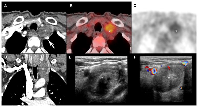
The vagus nerve (cranial nerve X) is the main nerve of the parasympathetic division of the autonomic nervous system. Vagal paragangliomas (VPGLs) are a prime example of an endocrine tumor associated with the vagus nerve. This rare, neural crest tumor constitutes the second most common site of hereditary head and neck paragangliomas (HNPGLs), most often in relation to mutations in the succinate dehydrogenase complex subunit D (SDHD) gene. The treatment paradigm for VPGL has progressively shifted from surgery to abstention or therapeutic radiation with curative-like outcomes. Parathyroid tissue and parathyroid adenoma can also be found in close association with the vagus nerve in intra or paravagal situations. Vagal parathyroid adenoma can be identified with preoperative imaging or suspected intraoperatively by experienced surgeons. Vagal parathyroid adenomas located in the neck or superior mediastinum can be removed via initial cervicotomy, while those located in the aortopulmonary window require a thoracic approach. This review particularly emphasizes the embryology, molecular genetics, and modern imaging of these tumors.

Keywords: diagnostic imaging; hyperparathyroidism; paragangliomas; vagus nerve.


Conflict of interest statement

Declaration of interest The authors declare that there is no conflict of interest that could be perceived as prejudicing the impartiality of the research reported.

Figures

Figure 1
Figure 1. Usual head and neck course of vagus nerve drawn on a cross sectional CT scanner with contrast injection
Vagus nerve (red spot) enters the lateral skull base in pars vascularis of jugular foramen (a) and the descends in the neck in the anterior angle formed by the posterolateral common carotid artery (CCA, white arrow) and posteromedial internal jugular vein (IJV, *) walls (b–e). In the upper thorax (f), vagal nerve is located anteriorly to sub-clavian artery (arrow heads).
Figure 2
Figure 2. Typical appearance of a VPGL on MRI
A: SE T1wi, B: SE T2wi, C: 3D Vibe T1wi with gadolinium and fat suppression, D: early arterial phase of a 2D T1 DCE) and 18F-FDOPA (E: PET/CT, F: PET). The VPGL is located in the left post-styloïd para-pharyngeal space (carotid space), splaying ICA (arrow head) and IJV (arrow).
Figure 3
Figure 3. Multifocal SDHD-related VPGL
Comparative cross sectional imaging centered on bilateral VPGL: 68Ga-DOTATATE (A, B), SE T2 wi (C), 3D gradient echo T1 wi with fat saturation (VIBE) after injection of gadolinium (D), Time of flight (TOF) MRA without gadolinium injection (E), early arterial phase of a 4D MRA with gadolinium injection. This case shows that VPGL (white arrows) were easily detected on 68Ga-DOTATATE PET/CT On MR, VPGL were not detected on conventional 2D T2, and hardly distinguishable on 3D T1 with gadolinium injection. AngioMR sequences confirm hyper-vascular pattern demonstrating high velocity arterial feeders on TOF (E) and early enhancement on 4D MRA with gadolinium injection. Note that on morphological data, these 2 mm VPGL were more easily detected on TOF sequence without injection. (Vessels are marked by a colour arrow, red: internal carotid artery, blue: internal jugular vein).
Figure 4
Figure 4. Sporadic VPGL
68Ga-DOTATATE (A, B), curved planar reconstructions of a 3D-GRE T1 Vibe with gadolinium and fat suppression (C) and 3D-SE (Space) T2 wi. An avid 68Ga-DOTATATE VPGL (*) is located at the level of mandibular angle. Adjacent to the VGPL 3D MRI demonstrates in this case the vagus nerve involvement due to its abnormal thickening. Abnormal thickening of the nerve is seen from lesion to the skull base (pars vascularis of jugular foramen) and may be due to vasa nervorum arterial feeders and edema more than peri-neural spread.
Figure 5
Figure 5. Schwannoma of the vagus nerve
Comparative cross sectional imaging: CT-scanner, arterial phase (A, D), 18FDG-PET (B, C), 2D ultrasound (E), 2D US with doppler (F). Multimodal assessment demonstrates a well-defined margin lesion (*) in cervico-thoracic region, located in the vagus nerve pathway, splaying right primitive carotid artery (arrow head) and right sub-clavian artery (arrow). This lesion does not enhance at the arterial phase CT (A, D), nor demonstrates hyper-vascularity at Doppler.
Figure 6
Figure 6. Cervical intravagal parathyroid adenoma
Axial CT with contrast injection (A) centered on the parathyroid adenoma (arrow) located in the vagus nerve path. Note the typical marked enhancement at arterial phase of injection of the parathyroid adenoma. Intraoperative view (B) showing the adenoma (arrow). In the present case, enucleation of the lesion, was not possible. Pathological analysis (C) revealed the presence of parathyroid tissue within the vagus nerve (*).
Figure 7
Figure 7. Vagal parathyroid adenoma located in the aortopulmonary window
4D-CT with non-enhanced (A), arterial (B) and delayed (C) phases, volume rendering of 4D-CT (arterial phase, D), Sestamibi SPECT acquisition (F). Parathyroid adenoma is located in the aortopulmonary window (arrow), under aortic arch (*) and above pulmonary artery (**). As for cervical localizations, this supernumerary ectopic parathyroid adenoma demonstrates avid enhancement (B, E) and rapid wash-out (C) compared to mediastinal lymph nodes (arrow heads). This adenoma was detected by sestamibi SPECT (F).

References

    1. al Zahrani A, Levine MA. Primary hyperparathyroidism. Lancet. 1997;349:1233–1238. - PubMed
    1. Archier A, Varoquaux A, Garrigue P, Montava M, Guerin C, Gabriel S, Beschmout E, Morange I, Fakhry N, Castinetti F, et al. Prospective comparison of (68)Ga-DOTATATE and (18)F-FDOPA PET/CT in patients with various pheochromocytomas and paragangliomas with emphasis on sporadic cases. European Journal of Nuclear Medicine and Molecular Imaging. 2016;43:1248–1257. - PubMed
    1. Arnault V, Beaulieu A, Lifante JC, Sitges Serra A, Sebag F, Mathonnet M, Hamy A, Meurisse M, Carnaille B, Kraimps JL. Multicenter study of 19 aortopulmonary window parathyroid tumors: the challenge of embryologic origin. World journal of surgery. 2010;34:2211–2216. - PubMed
    1. Arnold SM, Strecker R, Scheffler K, Spreer J, Schipper J, Neumann HP, Klisch J. Dynamic contrast enhancement of paragangliomas of the head and neck: evaluation with time-resolved 2D MR projection angiography. European radiology. 2003;13:1608–1611. - PubMed
    1. Baker CVH. The Embryology of Vagal Sensory Neurons. In: Undem BJ, Weinreich D, editors. Advances in Vagal Afferent Neurobiology. Boca Raton: CRC Press; 2005. pp. 3–26.

MeSH terms

LinkOut - more resources