. 2016 Jul 11:6:29703.
doi: 10.1038/srep29703.

An investigation of BMP-7 mediated alterations to BMP signalling components in human tenocyte-like cells

Affiliations

An investigation of BMP-7 mediated alterations to BMP signalling components in human tenocyte-like cells

Franka Klatte-Schulz et al. Sci Rep. .

Abstract

The incidence of tendon re-tears post-surgery is an ever present complication. It is suggested that the application of biological factors, such as bone morphogenetic protein 7 (BMP-7), can reduce complication rates by promoting tenogenic characteristics in in vitro studies. However, there remains a dearth of information in regards to the mechanisms of BMP-7 signalling in tenocytes. Using primary human tenocyte-like cells (hTLCs) from the supraspinatus tendon the BMP-7 signalling pathway was investigated: induction of the BMP associated Smad pathway and non-Smad pathways (AKT, p38, ERK1/2 and JNK); alterations in gene expression of BMP-7 associated receptors, Smad pathway components, Smad target gene (ID1) and tenogenic marker scleraxis. BMP-7 increases the expression of specific BMP associated receptors, BMPR-Ib and BMPR-II, and Smad8. Additionally, BMP-7 activates significantly Smad1/5/8 and slightly p38 pathways as indicated by an increase in phosphorylation and proven by inhibition experiments, where p-ERK1/2 and p-JNK pathways remain mainly unresponsive. Furthermore, BMP-7 increases the expression of the Smad target gene ID1, and the tendon specific transcription factor scleraxis. The study shows that tenocyte-like cells undergo primarily Smad8 and p38 signalling after BMP-7 stimulation. The up-regulation of tendon related marker genes and matrix proteins such as Smad8/9, scleraxis and collagen I might lead to positive effects of BMP-7 treatment for rotator cuff repair, without significant induction of osteogenic and chondrogenic markers.


Figures

Figure 1
Figure 1. Gene expression of BMP type I receptors in response to BMP-7.
Box plots represent qRT-PCR results given as mean normalised expression relative to 18S rRNA and the untreated control (reference line) using an efficiency corrected formula (n = 6). Symbols indicate statistically significant differences to the untreated control. (A) BMPR-Ia expression was increased after 2 to 8 h of BMP-7 stimulation (#p = 0.015, *p = 0.002). (B) BMPR-Ib expression was highly increased after 4 and 8 h of stimulation compared to unstimulated control (*p = 0.002). (C) ActR-I expression was increased at 2 h to 8 h with a peak at 4 h in the high BMP-7 group (*p = 0.002).
Figure 2
Figure 2. Gene expression of BMP type II receptors in response to BMP-7.
Box plots represent qRT-PCR results given as mean normalised expression relative to 18S rRNA and the untreated control (reference line) using an efficiency corrected formula. Symbols indicate significant differences to the untreated control (n = 6). (A) BMPR-II expression was strongly increased after 2 to 8 h of BMP-7 stimulation which was more pronounced in the high BMP-7 group (*p = 0.002. (B) The expression of ActR-IIa was increased in the low BMP-7 group after 1 h, in the high BMP-7 group after 2 h and 4 h after stimulation by both concentrations (#p = 0.03, *p = 0.004). (C) ActR-IIb expression was significantly down regulated after 2 h and 4 h compared to the untreated control (*p = 0.002).
Figure 3
Figure 3. Phosporylation of Smad1/5/8 after BMP-7 stimulation.
(A) Exemplary western blots show increased Smad 1/5/8 phosphorylation after 30 and 60 min of stimulation with BMP-7, but no increase in total Smad1 levels compared to unstimulated controls. GAPDH serves as reference protein. Imaging was conducted using the Odyssey imager and LiCor Odyssey software. (B) Quantification of relative phosphorylation normalised to total Smad1 and GAPDH was significantly increased at 30 and 60 min (n = 6). Stars mark significant differences to the untreated control, p = 0.002.
Figure 4
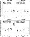
Figure 4. Gene expression of Smad1, 5 and 8/9 and co-Smad4.
Box plots represent qRT-PCR values given as mean normalised expression relative to 18S rRNA and the untreated control (reference line) using an efficiency corrected formula (n = 6). Stars indicate significant differences to the untreated control. (A) Expression of Smad1 was increased only at 4 h of BMP-7 stimulation (*p = 0.002). (B) Smad5 expression was not regulated compared to unstimulated control. (C) Smad8/9 expression increased after 4 h in the high BMP-7 group and highly increased after 8 h in both concentrations (*p = 0.002). (D) Smad4 expression was slightly increased after 1, 4 and 8 h of stimulation (*p = 0.002).
Figure 5
Figure 5. Phosphorylation of non-Smad signalling molecules.
(A) Exemplary western blots show increased p38 phosphorylation after 60 and 120 min. p-AKT was slightly increased after 120 min. No regulations were visible for p-Erk1/2 and p-JNK. Beta actin and GAPDH served as reference proteins. Imaging was conducted using the Odyssey imager and LiCor Odyssey software. (B) Quantification of 3 independent western blots (mean ± SD). Bands from the protein of interest were normalised to β-Actin or GAPDH and the unstimulated control, *p = 0.003, #p = 0.031, compared to control.
Figure 6
Figure 6. Gene expression of BMP target gene ID1 and tendon marker scleraxis.
Box plot graphs represent qRT-PCR values given as mean normalised expression relative to 18S rRNA and the untreated control (reference line) using an efficiency corrected formula (n = 6). Stars indicate significant differences to the untreated control. (A) ID1-expression was significantly increased at all concentrations and time points with the strongest increase at 8 h compared to the unstimulated control (*p = 0.002). (B) The expression of scleraxis was up-regulated at both concentrations for all time points and showed the strongest increase after 8 h of BMP-7 stimulation (*p = 0.002).
Figure 7
Figure 7. Inhibition of intracellular pathways, gene expression of BMP target gene ID1 and tendon marker scleraxis.
Box plot graphs represent qRT-PCR values given as mean normalised expression relative to 18S rRNA and the untreated control (reference line) using an efficiency corrected formula (n = 3). Inhibitors for the specific pathway were used in two concentrations. The inhibition of the Smad-pathway by LDN-193189 (LDN) resulted in a significant reduction of ID1 and scleraxis expression, less reduction was seen after inhibition of the p38 pathway with PD169316 (PD) and no effect after inhibition of the Akt-pathway (Akt), *p < 0.003, #p < 0.04, compared to BMP-7.
Figure 8
Figure 8. Gene expression of tenogenic, osteogenic, and chondrogenic markers to evaluate cell phenotype after BMP-7 stimulation.
Box plot graphs represent qRT-PCR values given as mean normalised expression relative to 18S rRNA and the untreated control (reference line) using an efficiency corrected formula (n = 6). Stars indicate significant differences to the untreated control. After 8 h of BMP-7-stimulation hTLCs expressed significantly higher amounts of the tenogenic markers scleraxis (p = 0.008) and matrix protein collagen I, whereas osteogenic markers were not upregulated or not expressed at all (Runx2 and osterix) and only Sox9 was upregulated as a marker for chondrogenic differentiation.

Similar articles

Cited by

References

    1. Aydin N., Kocaoglu B. & Guven O. “Single-row versus double-row arthroscopic rotator cuff repair in small- to medium-sized tears”, J. Shoulder. Elbow. Surg. 19(5), 722–725 (2010). - PubMed
    1. Gerhardt C. et al. “Arthroscopic single-row modified mason-allen repair versus double-row suture bridge reconstruction for supraspinatus tendon tears: a matched-pair analysis”, Am. J. Sports Med. 40(12), 2777–2785 (2012). - PubMed
    1. Klatte-Schulz F. et al. “Relationship between muscle fatty infiltration and the biological characteristics and stimulation potential of tenocytes from rotator cuff tears”, J. Orthop. Res. 32(1), 129–137 (2014). - PubMed
    1. Klatte-Schulz F. et al. “Characteristics and stimulation potential with BMP-2 and BMP-7 of tenocyte-like cells isolated from the rotator cuff of female donors”, PLoS. One. 8(6), e67209 (2013). - PMC - PubMed
    1. Klatte-Schulz F. et al. “Influence of age on the cell biological characteristics and the stimulation potential of male human tenocyte-like cells”, Eur. Cell Mater. 24, 74–89 (2012). - PubMed

Publication types

MeSH terms

Substances