. 2016 Jul 13:6:29316.
doi: 10.1038/srep29316.

Differential host susceptibility and bacterial virulence factors driving Klebsiella liver abscess in an ethnically diverse population

Affiliations

Differential host susceptibility and bacterial virulence factors driving Klebsiella liver abscess in an ethnically diverse population

I Russel Lee et al. Sci Rep. .

Abstract

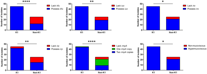
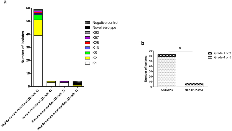
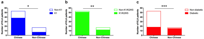
Hypervirulent Klebsiella pneumoniae is an emerging cause of community-acquired pyogenic liver abscess. First described in Asia, it is now increasingly recognized in Western countries, commonly afflicting those with Asian descent. This raises the question of genetic predisposition versus geospecific strain acquisition. We leveraged on the Antibiotics for Klebsiella Liver Abscess Syndrome Study (A-KLASS) clinical trial ongoing in ethnically diverse Singapore, to prospectively examine the profiles of 70 patients together with their isolates' genotypic and phenotypic characteristics. The majority of isolates belonged to capsule type K1, a genetically homogenous group corresponding to sequence-type 23. The remaining K2, K5, K16, K28, K57 and K63 isolates as well as two novel cps isolates were genetically heterogeneous. K1 isolates carried higher frequencies of virulence-associated genes including rmpA (regulator of mucoid phenotype A), kfu (Klebsiella ferric uptake transporter), iuc (aerobactin), iro (salmochelin) and irp (yersiniabactin) than non-K1 isolates. The Chinese in our patient cohort, mostly non-diabetic, had higher prevalence of K1 infection than the predominantly diabetic non-Chinese (Malays, Indian and Caucasian). This differential susceptibility to different capsule types among the various ethnic groups suggests patterns of transmission (e.g. environmental source, familial transmission) and/or genetic predisposition unique to each race despite being in the same geographical location.


Figures

Figure 1
Figure 1. K1 isolates carried higher frequencies of virulence-associated genes than non-K1 isolates.
These include kfu (100% vs 48%; P < 0.0001 by Fisher’s exact test), iuc (100% vs 76%; P = 0.001 by Fisher’s exact test), iro (100% vs 84%; P = 0.014 by Fisher’s exact test), irp (93.3% vs 60%; P = 0.001 by Fisher’s exact test) and rmpA (mean copy number: 1.98 vs 1.20; P < 0.0001 by Student’s t test). Expression of the hypermucoviscous phenotype correlated with possession of at least one rmpA copy in the genome.
Figure 2
Figure 2. K1/K2/K5 isolates were more resistant to serum exposure than non-K1/K2/K5 isolates.
(a) Serum resistance level of isolates from the different capsule types. Responses were graded as follows: grade 5 (highly serum-resistant), viable CFU after 3 hours of incubation in serum > 100% of the inoculum; grade 4 (serum-resistant), 71– 100%; grade 2 (serum-susceptible), 1–30%; grade 1 (highly serum-susceptible), 0%. The negative control, E. coli OP50, was highly-serum susceptible. (b) Comparison of prevalence of serum-resistant (grade 4 or 5) population between K1/K2/K5 isolates and non-K1/K2/K5 isolates (93.7% vs 57.1%; P = 0.019 using Fisher’s exact test).
Figure 3
Figure 3. The predominantly non-diabetic Chinese were more likely to be infected by K1 isolates and less likely to be infected by uncommon KLA capsule types (non-K1/K2/K5) than the predominantly diabetic non-Chinese.
(a) Comparison of prevalence of K1 infection between Chinese and non-Chinese (71.7% vs 41.2%; P = 0.040 by Fisher’s exact test). (b) Comparison of prevalence of non-K1/K2/K5 infection between Chinese and non-Chinese (3.8% vs 29.4%; P = 0.008 by Fisher’s exact test). (c) Comparison of prevalence of type 2 diabetes between Chinese and non-Chinese (34% vs 88.2%; P = 0.0001 by Fisher’s exact test).
Figure 4
Figure 4. The K1 strains SGH04 and NUH27 were more virulent than the non-K1 strain NUH29.
Bacterial burden in the organs of mice upon intraperitoneal injection with 104 or 105 CFUs of SGH04, NUH27 and NUH29. Each dot represents one infected mouse, whose liver, spleen and lungs were harvested 24 hours post-infection. Tissue homogenates that yielded no colonies were plotted with the value 10 CFU/g, which is the approximate limit of detection. The upper limit of quantification is 108 CFU/g.
Figure 5
Figure 5. Incidence of extraintestinal infection was not significantly different between obese/glucose intolerant mice and healthy mice upon oral inoculation with hvKP K1.
Data in (a–c) are means ± SEM. (a) Weight of mice fed a SCD or HFD over a course of 19 weeks. Ampicillin treatment between weeks 16 to 19 did not alter body mass. (b) Plasma glucose concentrations during the OGTT following 6 hours of fasting in mice fed a SCD or HFD for 16 weeks. Percentage change from basal (fasting glucose level at 0 min) in SCD-fed mice: 15 min = + 107.6% ± 10.0, 30 min = + 64.0% ± 6.3, 60 min = 26.7% ± 5.5, 120 min = + 1.1% ± 5.3. Percentage change from basal in HFD-fed mice: 15 min = + 121.2% ± 15.8, 30 min = + 88.8% ± 12.3, 60 min = 54.9% ± 8.8, 120 min = + 28.5% ± 5.4. Glucose elimination is significantly faster in SCD-fed than HFD-fed mice (P = 0.012 and 0.003 by Student’s t test, when comparing percentage change at 0–60 minutes and 0–120 minutes, respectively). (c) Area under the curve for glucose in (b), calculated using the trapezoidal rule. (d) Bacterial burden in the extraintestinal organs of SCD-fed (healthy) and HFD-fed (obese/glucose intolerant) mice that were treated with ampicillin or not, upon oral infection with 108 CFUs of the K1 strain SGH04. Each dot represents one infected mouse, whose liver and spleen were harvested 72 hours post-infection. Horizontal bars indicate geometric means. Tissue homogenates that yielded no colonies were plotted with the value 10 CFU/g, which is the approximate limit of detection.

References

    1. Shon A. S., Bajwa R. P. & Russo T. A. Hypervirulent (hypermucoviscous) Klebsiella pneumoniae: a new and dangerous breed. Virulence 4, 107–118, doi: 10.4161/viru.22718 (2013). - DOI - PMC - PubMed
    1. Siu L. K., Yeh K. M., Lin J. C., Fung C. P. & Chang F. Y. Klebsiella pneumoniae liver abscess: a new invasive syndrome. The Lancet Infectious Diseases 12, 881–887, doi: 10.1016/S1473-3099(12)70205-0 (2012). - DOI - PubMed
    1. Fang C. T. et al. Klebsiella pneumoniae genotype K1: an emerging pathogen that causes septic ocular or central nervous system complications from pyogenic liver abscess. Clinical Infectious Diseases 45, 284–293, doi: 10.1086/519262 (2007). - DOI - PubMed
    1. Fung C. P. et al. A global emerging disease of Klebsiella pneumoniae liver abscess: is serotype K1 an important factor for complicated endophthalmitis? Gut 50, 420–424 (2002). - PMC - PubMed
    1. Fang C. T., Chuang Y. P., Shun C. T., Chang S. C. & Wang J. T. A novel virulence gene in Klebsiella pneumoniae strains causing primary liver abscess and septic metastatic complications. The Journal of Experimental Medicine 199, 697–705, doi: 10.1084/jem.20030857 (2004). - DOI - PMC - PubMed

Publication types