Review
. 2016 Nov;100(5):979-984.
doi: 10.1189/jlb.4MR0316-102R. Epub 2016 Jul 12.

Angiogenesis and wound repair: when enough is enough

Affiliations
Review

Angiogenesis and wound repair: when enough is enough

Luisa A DiPietro. J Leukoc Biol. 2016 Nov.

Abstract

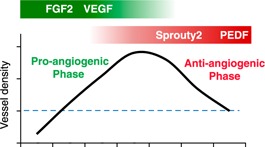
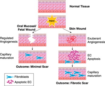
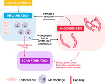
All animals heal, and the ability to heal is requisite for human health. One aspect of repair that has always been considered to be essential for adequate healing is the creation of a new vasculature via angiogenesis. As adult skin wounds heal, a period of rapid and robust capillary growth creates a vascular bed that has many fold more capillaries than does normal tissue. Over time, most of the newly formed capillaries regress, resulting in a final vascular density similar to that of normal skin. Certainly, new capillaries are necessary to bring nutrients, immune cells, and oxygen to healing wounds. Yet, the presumed functional importance of an overabundance of capillaries has recently been challenged, creating questions about whether excess capillary growth is truly necessary for healing. In particular, studies of wounds that heal exceptionally quickly and with less scar formation, such as those in fetal skin and oral mucosa, show that these tissues heal with a reduced angiogenic burst composed of more mature vessels that provide better oxygenation. The level of angiogenesis in wounds often correlates with the inflammatory response, largely because inflammatory cells produce an abundance of proangiogenic mediators. Both the selective reduction of inflammation and the selective reduction of angiogenesis have now been suggested as ways to improve scarring. These concepts link excessive inflammation and the production of a dense but poorly perfused capillary bed to inferior healing outcomes.

Keywords: VEGF; capillary; fibrosis; inflammation; scar; wound healing.


Figures

Figure 1
Figure 1
Regulation of wound angiogenesis in skin. During the healing of adult skin wounds, the number of capillaries increases dramatically to a level much greater than that in normal tissue. During the antiangiogenic phase, most of these newly formed vessels are pruned, creating a final vessel density that is similar to that of normal skin. Bars at the top of the graph show some critical proangiogenic (green) and antiangiogenic (red) signals. The blue dashed line indicates vessel density in normal tissue.
Figure 2
Figure 2
Comparison of angiogenesis in skin, oral mucosal, and fetal wounds. In skin, after an injury to the dermis and normal capillary bed occurs, a vigorous angiogenic response occurs (right). Eventually most of the new capillaries undergo apoptosis and capillary maturation results. In contrast, the angiogenic process in wounds of the oral mucosa and fetal skin (left) is limited, leading to more rapid capillary maturation. The differences in the angiogenic response have been linked to the final amount of scar formation. EC, endothelial cell.
Figure 3
Figure 3
Intersection of wound inflammation and angiogenesis with scar formation. Both inflammation and angiogenesis can directly influence scar formation (red arrows). During the acute inflammatory response, inflammatory cells may directly release profibrotic factors, notably TGF‐β. Independent of inflammation, the presence of a growing yet malformed vasculature that must eventually be removed can directly influence fibroblasts and scar outcomes, perhaps via edema, apoptosis, or the transition of recruited pericytes to an active fibroblast phenotype. Inflammation and angiogenesis are also intertwined as each process influences the other (black arrows). Macrophages, activated epithelial cells, and other inflammatory cells can release proangiogenic mediators such as VEGF and CXC chemokines, supporting robust capillary growth. In turn, the creation of the highly permeable temporary vasculature supports continued inflammation. Overall, inflammation and the immature capillary network can intersect to modulate scar formation. Each process can modulate the other, and each can independently influence scar formation in wounds. Dampening of either process, even independent of the other, has been shown to reduce scar formation.

References

    1. Gurtner, G. C. , Werner, S. , Barrandon, Y. , Longaker, M. T. (2008) Wound repair and regeneration. Nature 453, 314–321. - PubMed
    1. Eming, S. A. , Martin, P. , Tomic‐Canic, M. (2014) Wound repair and regeneration: mechanisms, signaling, and translation. Sci. Transl. Med. 6, 265sr6. - PMC - PubMed
    1. Eming, S. A. , Brachvogel, B. , Odorisio, T. , Koch, M. (2007) Regulation of angiogenesis: wound healing as a model. Prog. Histochem. Cytochem. 42, 115–170. - PubMed
    1. DiPietro, L. A. (2013) Angiogenesis and scar formation in healing wounds. Curr. Opin. Rheumatol. 25, 87–91. - PubMed
    1. Banda, M. J. , Knighton, D. R. , Hunt, T. K. , Werb, Z. (1982) Isolation of a nonmitogenic angiogenesis factor from wound fluid. Proc. Natl. Acad. Sci. USA 79, 7773–7777. - PMC - PubMed

Publication types

MeSH terms

Substances