. 2016 Jul 13:6:29760.
doi: 10.1038/srep29760.

Polyhydroxycurcuminoids but not curcumin upregulate neprilysin and can be applied to the prevention of Alzheimer's disease

Affiliations

Polyhydroxycurcuminoids but not curcumin upregulate neprilysin and can be applied to the prevention of Alzheimer's disease

Po-Ting Chen et al. Sci Rep. .

Abstract

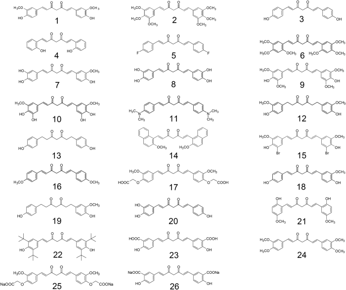
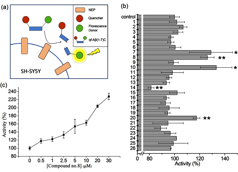
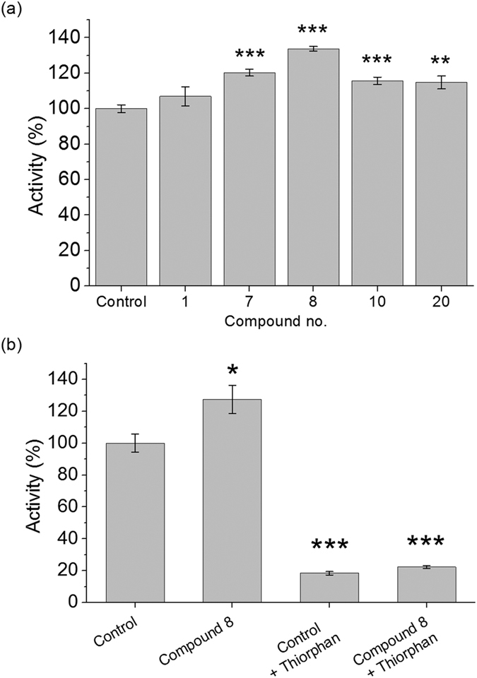
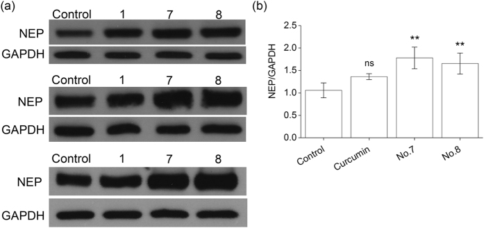
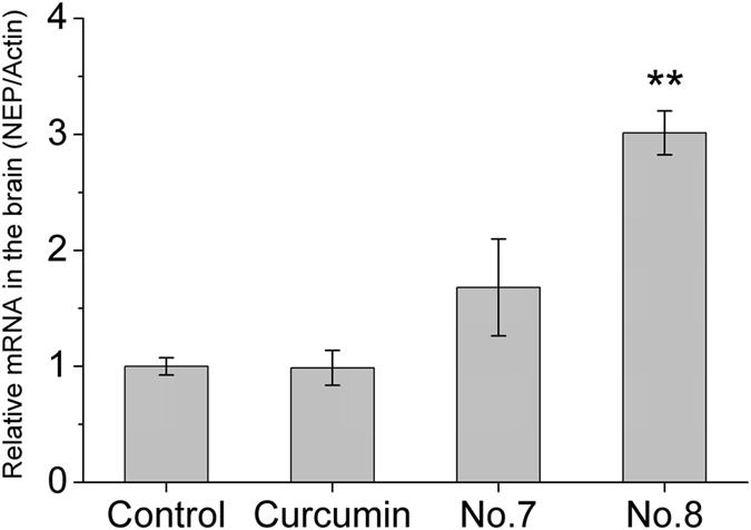
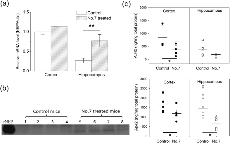
Neprilysin (NEP) is the most important Aβ-degrading enzyme. Its expression level decreases with age and inversely correlated with amyloid accumulation, suggesting its correlation with the late-onset of Alzheimer's disease. Recently, many reports showed that upregulating NEP level is a promising strategy in the prevention and therapy of Alzheimer's disease. Here, we used a sensitive fluorescence-based Aβ digestion assay to screen 25 curcumin analogs for their ability to upregulate NEP activity. To our surprise, four compounds, dihydroxylated curcumin, monohydroxylated demethoxycurcumin, and mono- and di-hydroxylated bisdemethoxycurcumin, increased NEP activity, while curcumin did not. The ability of these polyhydroxycurcuminoids to upregulate NEP was further confirmed by mRNA and protein expression levels in the cell and mouse models. Finally, feeding monohydroxylated demethoxycurcumin (also named demethylcurcumin) or dihydroxylated bisdemethoxycurcumin (also named bisdemethylcurcumin) to APPswe/PS1dE9 double transgenic mice upregulated NEP levels in the brain and reduced Aβ accumulation in the hippocampus and cortex. These polyhydroxycurcuminoids offer hope in the prevention of Alzheimer's disease.


Conflict of interest statement

There is potential competing financial interests. One US patent (US 9,074,238) has been granted.

Figures

Figure 1
Figure 1. Structures of curcumin (compound 1) and 25 curcumin analogs (compound 2-26).
1, 1,7-Bis(4-hydroxy-3-methoxyphenyl)-1,6-heptadiene-3,5-dione); 2, 1,7-Bis(3,4,5-trimethoxyphenyl)-1,6-heptadiene-3,5-dione; 3, 1,7-Bis(4-hydroxyphenyl)-1,6-heptadiene-3,5-dione; 4, 1,7-Bis(2-hydroxyphenyl)-1,6-heptadiene-3,5-dione; 5, 1,7-Bis(4-fluorophenyl)-1,6-heptadiene-3,5-dione; 6, 1,7-Bis(2,4,5-trimethoxyphenyl)-1,6-heptadiene-3,5-dione; 7, 1-(3,4-Dihydroxyphenyl)-7-(3-methoxy-4-hydroxyphenyl)-1,6-heptadiene-3,5-dione; 8, 1,7-Bis(3,4-dihydroxyphenyl)-1,6-heptadiene-3,5-dione; 9, 1,7-Bis(4-hydroxy-3,5-dimethoxyphenyl)-1,6-heptadiene-3,5-dione; 10, 1,7-Bis(3,4-dihydroxy-5-methoxyphenyl)-1,6-heptadiene-3,5-dione; 11, 1,7-Bis(4-N,N-dimethylaminophenyl)-1,6-heptadiene-3,5-dione; 12, 1,7-Bis(4-hydroxy-3-methoxyphenyl)heptan-3,5-dione; 13, 1,7-Bis(4-hydroxyphenyl)heptan-3,5-dione; 14, 1,7-Bis(2-methoxy-1-naphthyl)-1,6-heptadiene-3,5-dione; 15, 1,7-Bis(3-bromo-4-hydroxy-5-methoxyphenyl)-1,6-heptadiene-3,5-dione; 16, 1,7-Bis(4-methoxyphenyl)-1,6-heptadiene-3,5-dione; 17, 1,7-Bis(3-methoxy-4-carboxymethoxyphenyl)-1,6-heptadiene-3,5-dione; 18, 1-(4-Hydroxyphenyl)-7-(4-hydroxy-3-methoxyphenyl)-1,6-heptadiene-3,5-dione; 19, 1-(4-Hydroxyphenyl)-7-(4-hydroxy-3-methoxyphenyl)-heptan-3,5-dione; 20, 1-(3,4-Dihydroxyphenyl)-7-(4-hydroxyphenyl)-1,6-heptadiene-3,5-dione; 21, 1,7-Bis(2-hydroxy-5-methoxyphenyl)-1,6-heptadiene-3,5-dione; 22, 1,7-Bis(3,5-di-tert-butyl-4-hydroxyphenyl)-1,6-heptadiene-3,5-dione; 23, 1,7-bis(3-carboxy-4-hydroxyphenyl)-1,6-heptadiene-3,5-dione; 24, 1,7-Bis(3,4-dimethoxyphenyl)-1,6-heptadiene-3,5-dione; 25, 1,7-Bis(4-carboxymethoxy-3-methoxyphenyl)-1,6-heptadiene-3,5-dione disodium salt; 26, 1,7-bis(3-carboxy-4-hydroxyphenyl)-1,6-heptadiene-3,5-dione disodium salt.
Figure 2
Figure 2. Screen compounds with the ability to increase Aβ-degradation.
(a) Aβ-degrading activity assay using qf-Aβ(1-7)C as substrate. (b) Results for SH-SY5Y cells incubated for 24 h with 0.5% DMSO (control) or the compound listed on the y axis (5 μM). (c) Aβ-degrading activity assay using different concentrations of compound 8. In (b,c), the data are presented as the mean ± SD for three independent samples; in (b) *p < 0.05; **p < 0.01 compared to the control by one-way ANOVA.
Figure 3
Figure 3. Examination of the increased Aβ-degrading activity coming from the action of NEP.
(a) Aβ-degrading activity assay using qf-Aβ(12-16)AAC as substrate. SH-SY5Y cells were incubated 12 h with 0.5% DMSO (control) or the indicated compounds (5 μM) before assay. (b) Thiorphan inhibition assay. SH-SY5Y cells were incubated for 12 h with 0.5% DMSO or compound 8 (5 μM), then with or without 50 μM thiorphan for 30 min before Aβ-degrading activity was measured. The data are the mean ± SD for three independent samples. *p < 0.05; **p < 0.01; ***p < 0.001 compared to the control by Student’s t-test.
Figure 4
Figure 4. Western blot analysis and quantitative densitometry for NEP levels in RA-differentiated SH-SY5Y cells.
The cells were incubated with 0.5% DMSO (control) or 5 μM curcumin (no. 1) or compound 7 or 8 for 24 h, then NEP protein in the cell membrane fraction was measured by western blotting. (a) Western blotting results of three independent experiments. GAPDH was used as the loading control. (b) The quantification of NEP protein levels by Image J. The NEP intensities were normalized to the GAPDH intensities for three independent experiments. Data are presented as the mean ± SD; ns, not significant; **p < 0.01 compared to the DMSO-treated control group by Student’s t-test.
Figure 5
Figure 5. NEP mRNA levels in the brains of mice fed vehicle (control) or the indicated compound for 7 days (10 mg/kg BW per day) by gavage (n = 5).
β-actin was used as the reference gene. The results are the mean ± SEM; **p < 0.01 compared to the vehicle-fed group by Student’s t-test.
Figure 6
Figure 6. Effect of compound 7 on NEP mRNA and protein levels and Aβ clearance in vivo.
APPswe/PS1dE9 mice were fed vehicle (control) or compound 7 (10 mg/kg BW) six times per week for 6.5 months by gavage. (a) NEP mRNA levels in the cortex and hippocampus using β-actin as the reference gene, with the value for the cortex of the vehicle-fed control group set as 1. The results are presented as the mean ± SEM (n = 5). (b) Neprilysin western blot. Protein samples of 12 μg extracted from the hippocampus of eight APPswe/PS1dE9 mice fed with vehicle or compound 7 were resolved on an 8% Bis-Tris gel and immunoblotted with rat monoclonal anti-mouse NEP antibody. (c) ELISA results for formic acid-extracted (insoluble) Aβ40 (top panel) and Aβ42 (bottom panel) in the cortex and hippocampus; the data for each mouse are indicated and the mean is indicated by the line (n = 5). The heavy black line indicates the sets of data showing a significant difference; *p < 0.05; **p < 0.01 compared to the control by Student’s t-test.
Figure 7
Figure 7. Effect of compound 8 on reducing amyloid plaque burden in the mouse brain.
The APPswe/PS1dE9 mice were treated with vehicle or compound 8 for 8 months. Plaque burden of the 11-month-old WT littermate (n = 4), vehicle-treated (n = 3) and compound 8-treated transgenic mice (n = 6) were analyzed by ThS staining (4 slices per mouse). (a) Representative ThS-staining images. (b,c) Plaque number and percentage of plaque-covered area in the cortex (b) and hippocampus (c) were quantitated by ImageJ. Data was expressed in mean ± SEM (*p < 0.05, **p < 0.01, ***p < 0.001, ****p < 0.0001 by one-way ANOVA with Fisher’s LSD test).
Figure 8
Figure 8. Effects of curcumin and compounds 7 and 8 on DNA methylation of NEP in the N2a cells.
The genomic structure of the CpG island in the mouse NEP promoter region is drawn to scale above the bisulfite sequencing chromatogram. The CpG sites are indicated by number. Each row of circles indicates a single cloned allele. Black circle, methylated cytosine; white circle, unmethylated cytosine.

References

    1. Lannfelt L. et al.. Perspectives on future Alzheimer therapies: amyloid-beta protofibrils - a new target for immunotherapy with BAN2401 in Alzheimer’s disease. Alzheimers Res. Ther. 6, 16 (2014). - PMC - PubMed
    1. Moreth J., Mavoungou C. & Schindowski K. Passive anti-amyloid immunotherapy in Alzheimer’s disease: What are the most promising targets? Immun. Ageing 10, 18 (2013). - PMC - PubMed
    1. Iwata N., Higuchi M. & Saido T. C. Metabolism of amyloid-b peptide and Alzheimer’s disease. Pharmacol. Therapeut. 108, 129–148 (2005). - PubMed
    1. Wang D. S., Iwata N., Hama E., Saido T. C. & Dickson D. W. Oxidized neprilysin in aging and Alzheimer’s disease brains. Biochem. Biophys. Res. Commun. 310, 236–241 (2003). - PubMed
    1. Hellstrom-Lindahl E., Ravid R. & Nordberg A. Age-dependent decline of neprilysin in Alzheimer’s disease and normal brain: inverse correlation with A beta levels. Neurobiol. Aging 29, 210–221 (2008). - PubMed