

Clinical Applications of PET and PET-CT
- PMID: 27408291
- PMCID: PMC4921358
- DOI: 10.1016/S0377-1237(09)80099-3
Clinical Applications of PET and PET-CT
Abstract
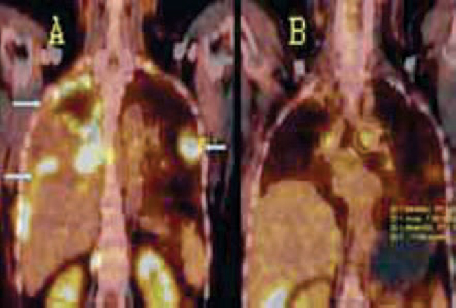
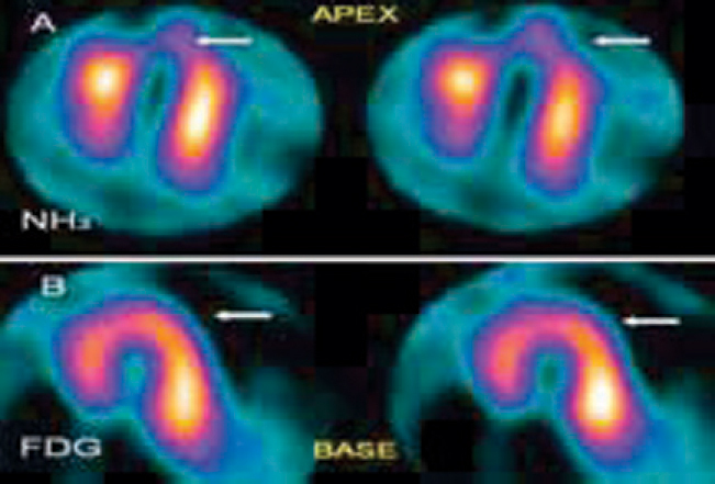
Positron emission tomography (PET) and PET/ computed tomography (CT) are emerging as important imaging techniques and their popularity is growing within the medical fraternity. Though PET has been a useful research tool for many decades its real growth into clinical applications has occurred in the last one decade or so. Currently its major use is in oncologic imaging. However it has a multitude of clinical applications in cardiology, neurology and psychiatry as well. In oncologic imaging, a major advantage of PET is that a single whole-body examination can provide accurate assessment of disease activity and spread. PET/CT amalgamates the functional information of PET with the structural details of the CT scan, thus greatly aiding in accurate staging, therapy response assessment and early detection of recurrent disease.
Keywords: Positron emission tomography.
Figures
References
-
- Coleman RE, Delbeke D, Guiberteau MJ. Concurrent PET/CT with an integrated imaging System: Intersociety Dialogue from the Joint Working Group of the American College of Radiology, the society of nuclear medicine and the society of computed body tomography and magnetic resonance. J Nucl Med. 2005;46:1225–1239. - PubMed
-
- Jadvar H, Parker JA. In Clinical PET and PET/CT. first ed. Springer; Philadelphia: 2005. PET radiotracers; pp. 45–67.
-
- Zeissmann HA, O'Malley JP, Thrall JH. Third ed. Elsevier; London: 2006. In Nuclear Medicine the requisites.
-
- Gambhir SS, Czernin J, Schwimmer J, Silverman DH, Coleman RE, Phelps ME. A tabulated summary of the FDG PET literature. J Nucl Med. 2001;42:1S–93S. - PubMed
-
- Ciernik IF, Dizendorf E, Baumert BG. Radiation treatment planning with an integrated positron emission and computer tomography (PET/CT): a feasibility study. Int J Radiat Oncol Biol Phys. 2003;57:853–863. - PubMed
LinkOut - more resources
Full Text Sources
Other Literature Sources
