Review
. 2016 Jun 27:5:F1000 Faculty Rev-1513.
doi: 10.12688/f1000research.8206.1. eCollection 2016.

Pathogenesis of myasthenia gravis: update on disease types, models, and mechanisms

Affiliations
Review

Pathogenesis of myasthenia gravis: update on disease types, models, and mechanisms

William D Phillips et al. F1000Res. .

Abstract

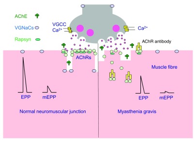
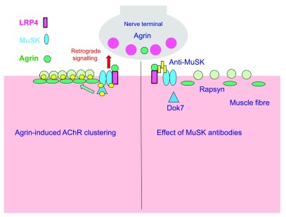
Myasthenia gravis is an autoimmune disease of the neuromuscular junction (NMJ) caused by antibodies that attack components of the postsynaptic membrane, impair neuromuscular transmission, and lead to weakness and fatigue of skeletal muscle. This can be generalised or localised to certain muscle groups, and involvement of the bulbar and respiratory muscles can be life threatening. The pathogenesis of myasthenia gravis depends upon the target and isotype of the autoantibodies. Most cases are caused by immunoglobulin (Ig)G1 and IgG3 antibodies to the acetylcholine receptor (AChR). They produce complement-mediated damage and increase the rate of AChR turnover, both mechanisms causing loss of AChR from the postsynaptic membrane. The thymus gland is involved in many patients, and there are experimental and genetic approaches to understand the failure of immune tolerance to the AChR. In a proportion of those patients without AChR antibodies, antibodies to muscle-specific kinase (MuSK), or related proteins such as agrin and low-density lipoprotein receptor-related protein 4 (LRP4), are present. MuSK antibodies are predominantly IgG4 and cause disassembly of the neuromuscular junction by disrupting the physiological function of MuSK in synapse maintenance and adaptation. Here we discuss how knowledge of neuromuscular junction structure and function has fed into understanding the mechanisms of AChR and MuSK antibodies. Myasthenia gravis remains a paradigm for autoantibody-mediated conditions and these observations show how much there is still to learn about synaptic function and pathological mechanisms.

Keywords: AChR; Myasthenia gravis; immunoglobulin; neuromuscular junction.


Conflict of interest statement

Competing interests: The University of Oxford holds a patent for MuSK antibody detection, licensed in the USA to Athena Diagnostics, and Angela Vincent receives a proportion of royalties. William D. Phillips has no disclosures.

No competing interests were disclosed.

Figures

Figure 1.
Figure 1.. Assessing neuromuscular transmission.
( A) Healthy neuromuscular transmission. The nerve terminal can release the contents of each vesicle (quanta) of acetylcholine by exocytosis. Spontaneous release of single quanta of acetylcholine activates the intrinsic cation channels of acetylcholine receptors (AChRs) in the postsynaptic membrane to produce a small, transient depolarisation called a miniature endplate potential (mEPP). The nerve action potential opens voltage-gated calcium channels (VGCCs) and triggers exocytosis of many quanta of acetylcholine, simultaneously producing the (much larger) EPP. In healthy individuals, the amplitude of the EPP is more than enough to reach the threshold required to activate the postsynaptic voltage-gated sodium channels (VGNaCs) and generate a muscle action potential. ( B) The myasthenia gravis neuromuscular junction. AChR antibodies (mainly immunoglobulin [Ig]G1) activate complement, resulting in membrane attack complex-mediated damage to the post-junctional membrane architecture. The postsynaptic AChR numbers are depleted by divalent antibodies inducing AChR internalisation. The loss of AChRs results in smaller mEPP and EPP amplitudes. The EPP may not reach threshold, especially when the nerve is repetitively activated. Abbreviations: AChE, acetylcholinesterase
Figure 2.
Figure 2.. Disruption of postsynaptic differentiation pathway by muscle-specific kinase (MuSK) autoantibodies.
( A) Healthy MuSK-mediated postsynaptic differentiation pathway at the neuromuscular junction (NMJ). Neural agrin secreted by the motor nerve terminal binds to LRP4, low-density lipoprotein receptor-related protein 4 (LRP4), which causes the dimerisation of MuSK. MuSK dimerisation causes phosphorylation of MuSK and associated proteins of the MuSK pathway, including Dok7 and the acetylcholine receptor (AChR) β-subunit. Rapsyn is recruited to the phosphorylated AChRs, stabilising postsynaptic clusters of AChRs. ( B) Impaired postsynaptic differentiation in animal models of MuSK myasthenia gravis. MuSK autoantibodies are mainly of the immunoglobulin (Ig)G4 subclass. They block the assembly of the agrin-LRP4-MuSK complex. Interruption of MuSK kinase signalling leads to slow disassembly of the postsynaptic AChR clusters. A resultant decline in miniature endplate potential (mEPP) and EPP amplitude (not shown) results in failure of the muscle action potential and fatiguing weakness. Co-existing IgG1-3 antibodies, although lower concentration, may contribute but their pathogenic roles are not yet well defined. The compensatory presynaptic upregulation of quantal release found in AChR MG does not occur in MuSK MG.

References

    1. Berrih-Aknin S, Frenkian-Cuvelier M, Eymard B: Diagnostic and clinical classification of autoimmune myasthenia gravis. J Autoimmun. 2014;48–49:143–148. 10.1016/j.jaut.2014.01.003 - DOI - PubMed
    1. Vincent A: Unravelling the pathogenesis of myasthenia gravis. Nat Rev Immunol. 2002;2(10):797–804. 10.1038/nri916 - DOI - PubMed
    1. Verschuuren JJ, Huijbers MG, Plomp JJ, et al. : Pathophysiology of myasthenia gravis with antibodies to the acetylcholine receptor, muscle-specific kinase and low-density lipoprotein receptor-related protein 4. Autoimmun Rev. 2013;12(9):918–923. 10.1016/j.autrev.2013.03.001 - DOI - PubMed
    1. Phillips WD, Christadoss P, Losen M, et al. : Guidelines for pre-clinical animal and cellular models of MuSK-myasthenia gravis. Exp Neurol. 2015;270:29–40. 10.1016/j.expneurol.2014.12.013 - DOI - PubMed
    1. Plomp JJ, Morsch M, Phillips WD, et al. : Electrophysiological analysis of neuromuscular synaptic function in myasthenia gravis patients and animal models. Exp Neurol. 2015;270:41–54. 10.1016/j.expneurol.2015.01.007 - DOI - PubMed