

Incarcerated ovarian herniation of the canal of Nuck in a female infant: Ultrasonographic findings and review of literature
- PMID: 27408712
- PMCID: PMC4925906
- DOI: 10.1016/j.amsu.2016.06.003
Incarcerated ovarian herniation of the canal of Nuck in a female infant: Ultrasonographic findings and review of literature
Abstract
Introduction: Inguinal hernia with containing the ovary presenting as a palpable groin mass is an uncommon congenital condition, and it may cause complications such as strangulation, torsion, and infertility. We present a case of ovarian herniation into inguinal canal with sonographic findings.
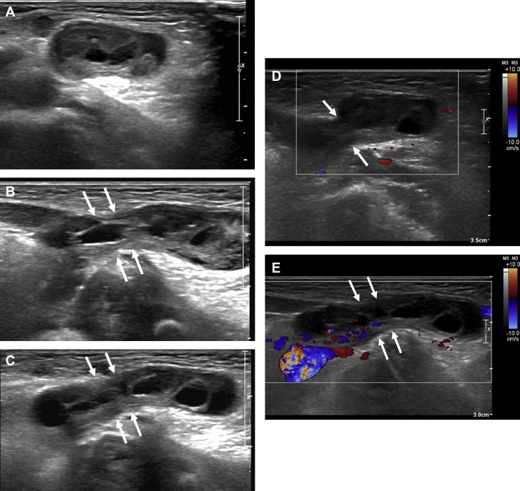
Presentation of case: A 15-day-old infant girl visited our hospital with the complaints of palpable mass in the right groin. On physical examination, a palpable non-movable mass was found in the right inguinal region, and it was irreducible. During sonographic examination, a well-circumscribed solid mass containing small cysts was found. Then, oophorectomy with high inguinal ligation was performed, and the patient was doing well after surgery.
Discussion: The canal of Nuck is an abnormal patent pouch of the parietal peritoneum extending to the round ligament of the uterus into the labia majora through the inguinal ring. When this canal obliterates incompletely, inguinal herniation of ovary or hydrocele occur in the female children. In the clinical practice, ovarian herniation should be differentiated from a hernia containing intestine, fat, fluid, or lymph nodes. Therefore, a careful sonographic evaluation is mandatory to make an accurate diagnosis in female infants with palpable inguinal mass.
Conclusion: Ultrasound (US) with color Doppler US can be helpful to the diagnosis of ovarian herniation through the canal of Nuck and hernia-related complications.
Keywords: Canal of Nuck; Inguinal hernia; Ovary; Ultrasound.
Figures
References
-
- George E.K., Oudesluys-Murphy A.M., Madern G.C., Cleyndert P., Blomjous J.G. Inguinal hernias containing the uterus, fallopian tube, and ovary in premature female infants. J. Pediatr. 2000;136:696–698. - PubMed
-
- Bronsther B., Abrams M.W., Elboim C. Inguinal hernia in children: a study of 1000 cases and a review of the literature. J. Am. Med. Womens Assoc. 1972;27:522–535. - PubMed
-
- Yigit H., Tuncbilek I., Fitoz S., Yigit N., Kosar U., Karabulut B. Cyst of the canal of Nuck with demonstration of the proximal canal: the role of the compression technique in sonographic diagnosis. J. Ultrasound Med. 2006;25:123–125. - PubMed
-
- Park S.J., Lee H.K., Hong H.S., Kim H.C., Kim D.H., Park J.S. Hydrocele of the canal of Nuck in a girl: ultrasound and MR appearance. Br. J. Radiol. 2004;77:243–244. - PubMed
-
- Ming Y.C., Luo C.C., Chao H.C., Chu S.M. Inguinal hernia containing uterus and uterine adnexa in female infants: report of two cases. Pediatr. Neonatol. 2011;52:103–105. - PubMed
Publication types
LinkOut - more resources
Full Text Sources
Other Literature Sources
Research Materials
