Review
. 2015 Jun 17:3:18.
doi: 10.1186/s40560-015-0083-6. eCollection 2015.

The role of spontaneous effort during mechanical ventilation: normal lung versus injured lung

Affiliations
Review

The role of spontaneous effort during mechanical ventilation: normal lung versus injured lung

Takeshi Yoshida et al. J Intensive Care. .

Abstract

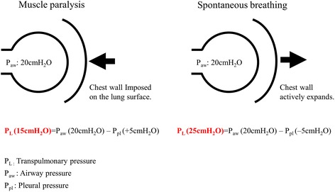
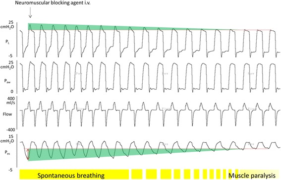
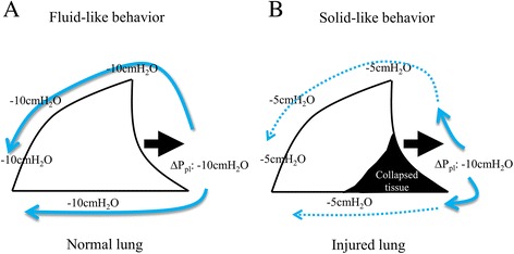
The role of preserving spontaneous effort during mechanical ventilation and its interaction with mechanical ventilation have been actively investigated for several decades. Inspiratory muscle activities can lower the pleural components surrounding the lung, leading to an increase in transpulmonary pressure when spontaneous breathing effort is preserved during mechanical ventilation. Thus, increased transpulmonary pressure provides various benefits for gas exchange, ventilation pattern, and lung aeration. However, it is important to note that these beneficial effects of preserved spontaneous effort have been demonstrated only when spontaneous effort is modest and lung injury is less severe. Recent studies have revealed the 'dark side' of spontaneous effort during mechanical ventilation, especially in severe lung injury. The 'dark side' refers to uncontrollable transpulmonary pressure due to combined high inspiratory pressure with excessive spontaneous effort and the injurious lung inflation pattern of Pendelluft (i.e., the translocation of air from nondependent lung regions to dependent lung regions). Thus, during the early stages of severe ARDS, the strict control of transpulmonary pressure and prevention of Pendelluft should be achieved with the short-term use of muscle paralysis. When there is preserved spontaneous effort in ARDS, spontaneous effort should be maintained at a modest level, as the transpulmonary pressure and the effect size of Pendelluft depend on the intensity of the spontaneous effort.

Keywords: ARDS; Lung injury; Muscle paralysis; Pendelluft; Pleural pressure; Spontaneous breathing; Transpulmonary pressure.


Figures

Figure 1
Figure 1
Transpulmonary pressure difference: muscle paralysis vs. spontaneous breathing. Diaphragmatic contraction can elevate transpulmonary pressure with the same airway pressure applied in muscle paralysis, by altering the pleural components surrounding the lung.
Figure 2
Figure 2
Transition phase from spontaneous breathing to muscle paralysis in a rabbit. A lung-injured animal was ventilated with assisted pressure control mode. We recorded continuously waveforms of transpulmonary pressure, airway pressure, flow, and esophageal pressure without any change in ventilatory settings, after injection of neuromuscular blocking agent. When spontaneous breathing during mechanical ventilation is diminished, the negative swing in esophageal pressure is decreasing. As a result, inspiratory transpulmonary pressure decreases. Note that inspiratory transpulmonary pressure linearly correlates with the intensity of spontaneous breathing effort.
Figure 3
Figure 3
Fluid-like behavior presented in normal lung vs. solid-like behavior presented in injured lung. (A) The normal lung is traditionally considered to be a continuous elastic system—exhibiting fluid-like behavior—such that distending pressure applied to a local region of the pleura (the negative swing in pleural pressure generated by diaphragmatic contraction is −10 cm H2O) becomes generalized over the whole lung (pleural) surface (the negative swings in pleural pressure at any regions are the same −10 cm H2O). (B) In injured lung, the negative swing in pleural pressure generated by diaphragmatic contraction is not uniformly transmitted, but rather concentrated in the dependent lung regions, thus a huge difference in negative pleural pressure between nondependent and dependent lung regions was generated at the early phase of inspiration, causing Pendelluft. Adapted with permission of the Wolters Kluwer Health (Ref. [36]).
Figure 4
Figure 4
EIT waveforms in experimental lung injury—spontaneous versus mechanical breaths. Note that the early inflation in the dependent region (Zones 3 and 4) was accompanied by concomitant deflation of nondependent region (Zones 1 and 2), indicating movement of air from nondependent to dependent lung (i.e. Pendelluft). Note that under the same tidal volume, spontaneous breathing during mechanical ventilation unsuspectedly increased dependent lung inflation (Zones 3 and 4) due to Pendelluft. Adapted with permission of the American Thoracic Society Copyright © 2014 American Thoracic Society (Ref. [12]).
Figure 5
Figure 5
CT images in experimental lung injury—muscle paralysis vs. spontaneous breathing. Both dynamic CT images of the same anatomical, sagittal level at end-expiration are shown to compare the end-expiratory lung volume and the shape of the diaphragm. CT images are colored according to their Hounsfield units densities. The black lines indicate the diaphragm at end-expiration. These dynamic CT images were continuously taken after injection of neuromuscular blocking agent, without any change in ventilatory settings. Spontaneous breathing effort restored the end-expiratory lung volume due to diaphragmatic muscle tone. Once diaphragm was paralyzed, diaphragm shifted to cranial direction, resulting in large collapse in dorsal lung regions. Note that this happened because inadequate (low) PEEP was applied.

References

    1. Stock MC, Downs JB, Frolicher DA. Airway pressure release ventilation. Crit Care Med. 1987;15:462–6. doi: 10.1097/00003246-198705000-00002. - DOI - PubMed
    1. Putensen C, Zech S, Wrigge H, Zinserling J, Stüber F, Von Spiegel T, et al. Long-term effects of spontaneous breathing during ventilatory support in patients with acute lung injury. Am J Respir Crit Care Med. 2001;164:43–9. doi: 10.1164/ajrccm.164.1.2001078. - DOI - PubMed
    1. Marini JJ. Spontaneously regulated vs. controlled ventilation of acute lung injury/acute respiratory distress syndrome. Curr Opin Crit Care. 2011;17:24–9. doi: 10.1097/MCC.0b013e328342726e. - DOI - PubMed
    1. Putensen C, Muders T, Varelmann D, Wrigge H. The impact of spontaneous breathing during mechanical ventilation. Curr Opin Crit Care. 2006;12:13–8. doi: 10.1097/01.ccx.0000198994.37319.60. - DOI - PubMed
    1. Wrigge H, Zinserling J, Neumann P, Defosse J, Magnusson A, Putensen C, et al. Spontaneous breathing improves lung aeration in oleic acid-induced lung injury. Anesthesiology. 2003;99:376–84. doi: 10.1097/00000542-200308000-00019. - DOI - PubMed

LinkOut - more resources