. 2016 May 3;5(7):449-458.
doi: 10.1016/j.molmet.2016.04.007. eCollection 2016 Jul.

Comprehensive alpha, beta and delta cell transcriptomes reveal that ghrelin selectively activates delta cells and promotes somatostatin release from pancreatic islets

Affiliations

Comprehensive alpha, beta and delta cell transcriptomes reveal that ghrelin selectively activates delta cells and promotes somatostatin release from pancreatic islets

Michael R DiGruccio et al. Mol Metab. .

Abstract

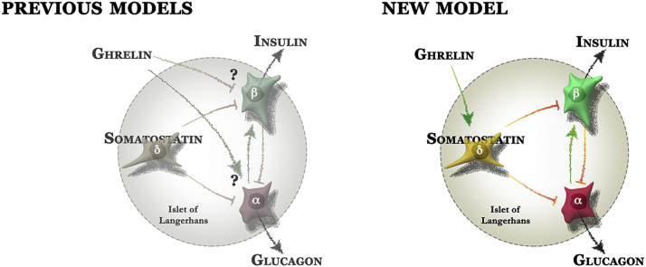
Objective: Complex local crosstalk amongst endocrine cells within the islet ensures tight coordination of their endocrine output. This is illustrated by the recent demonstration that the negative feedback control by delta cells within pancreatic islets determines the homeostatic set-point for plasma glucose during mouse postnatal development. However, the close association of islet endocrine cells that facilitates paracrine crosstalk also complicates the distinction between effects mediated directly on beta cells from indirect effects mediated via local intermediates, such as somatostatin from delta cells.

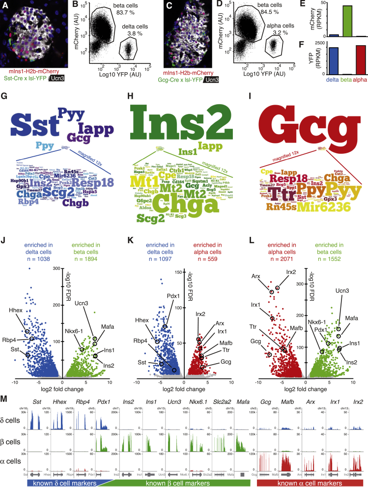
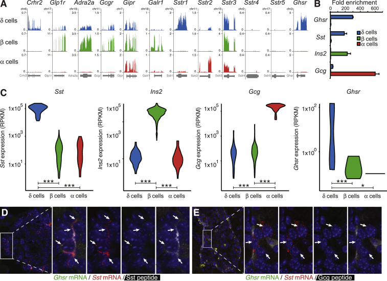
Methods: To resolve this problem, we generated reporter mice that allow collection of pure pancreatic delta cells along with alpha and beta cells from the same islets and generated comprehensive transcriptomes for each islet endocrine cell type. These transcriptomes afford an unparalleled view of the receptors expressed by delta, alpha and beta cells, and allow the prediction of which signal targets which endocrine cell type with great accuracy.

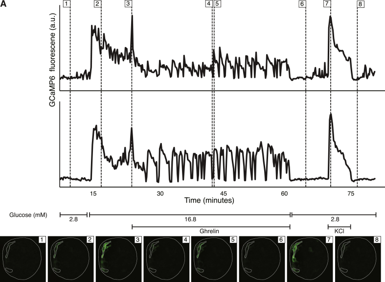
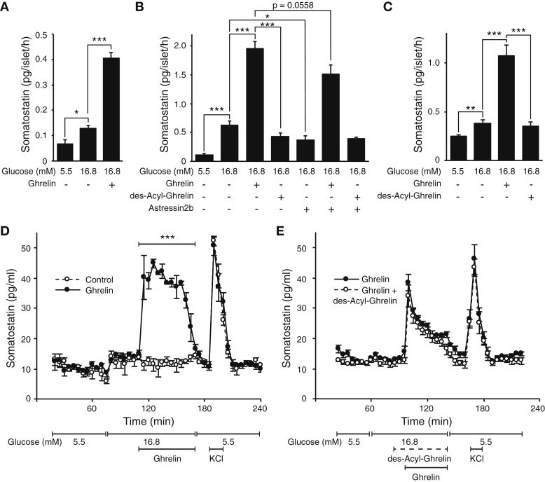
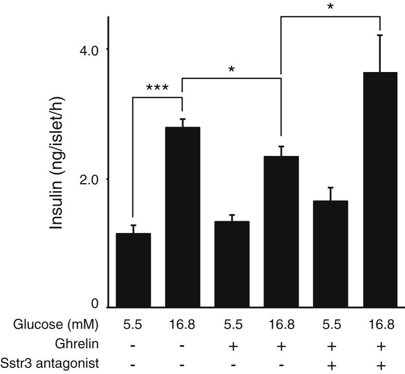
Results: From these transcriptomes, we discovered that the ghrelin receptor is expressed exclusively by delta cells within the islet, which was confirmed by fluorescent in situ hybridization and qPCR. Indeed, ghrelin increases intracellular calcium in delta cells in intact mouse islets, measured by GCaMP6 and robustly potentiates glucose-stimulated somatostatin secretion on mouse and human islets in both static and perfusion assays. In contrast, des-acyl-ghrelin at the same dose had no effect on somatostatin secretion and did not block the actions of ghrelin.

Conclusions: These results offer a straightforward explanation for the well-known insulinostatic actions of ghrelin. Rather than engaging beta cells directly, ghrelin engages delta cells to promote local inhibitory feedback that attenuates insulin release. These findings illustrate the power of our approach to resolve some of the long-standing conundrums with regard to the rich feedback that occurs within the islet that is integral to islet physiology and therefore highly relevant to diabetes.

Keywords: Alpha cell; Beta cell; Crhr2, Corticotropin-releasing hormone receptor type 2; Delta cell; FISH, Fluorescent in situ hybridization; GSSS, Glucose-stimulated somatostatin secretion; Ghrelin; Ghsr, Growth hormone secretagogue receptor; Iapp, Islet amyloid polypeptide; RPKM, Reads per kilobase gene model per million reads sequenced; Somatostatin release; Transcriptome; Trpm2, Transient receptor potential melastatin 2; Ucn3, Urocortin 3; YFP, Yellow fluorescent protein.


Figures

None
Graphical abstract
Figure 1
Figure 1
Generation and validation of the delta, beta, and alpha cell transcriptomes. A: Islet of a mIns1-H2b-mCherry × Sst-Cre × LSL-YFP triple transgenic mouse labels all beta cells with nuclear mCherry (counterstained in white with Ucn3) and labels all delta with YFP (stained in green). B: FACS plots of dissociated beta and delta cells from this same cross. C: Islet of a mIns1-H2b-mCherry × Gcg-Cre × LSL-YFP triple transgenic mouse labels all beta cells with nuclear mCherry (counterstained in white with Ucn3) and labels alpha cells with YFP (stained in green). D: FACS plots of dissociated alpha and delta cells from this same cross. E, F: Beta cell transcriptomes are highly enriched for mCherry reads while delta and alpha cell transcriptomes are highly enriched for YFP reads, confirming our transgenic purification strategy. RPKM = reads per kilobase gene model per million reads sequenced. G–I: Graphical representation of the relative expression of the 100 most-abundantly detected transcripts in each cell type. Note that most of these genes are expressed at levels that are so much lower compared to the most abundant transcript in each endocrine cell type that they were magnified 12x to maintain legibility. J–L: Volcano plots listing the number of significantly enriched genes for each pairwise comparison. Selected markers of alpha, beta, and delta cell identity are highlighted for visual reference. M: Genome browser plots comparing the expression of a large panel of markers with well-established expression patterns validate the high degree of purity achieved in these delta, beta, and alpha cells transcriptomes.
Figure 2
Figure 2
Delta cells selectively express Ghsr. A: Normalized browser plots illustrating the expression of a series of GPCRs in delta, beta and alpha cells of the mouse islet. B: Confirmation by qPCR that Ghsr message is selectively expressed by pancreatic delta cells. C: Violin plots of single cell RNA-seq of wild type mouse pancreatic islet cells confirms that Ghsr expression is detectable only in delta cells. D: FISH confirmation of the expression of Ghsr gene (green dots) in pancreatic delta cells of wild type mice, colocalized with Sst message (red dots) and Sst peptide (white). E: mRNA for Ghsr (green dots) and Sst (red dots) co-localizes in a peripheral islet population that does not express Gcg peptide (white). *P < 0.05; **P < 0.01; ***P < 0.001.
Figure 3
Figure 3
Ghrelin activates an intracellular calcium response as measured by GCaMP6 in intact islets. A: Calcium responses were measured over time in intact islets using the genetically encoded calcium sensor GCaMP6 expressed selectively in delta cells (Sst-Cre × LSL-GCaMP6). Calcium traces of two representative cells in response to changes in glucose concentration and stimulation with 100 nM ghrelin or 30 mM KCl, as indicated. Thumbnails of the islet at the indicated time points in each graph are given, with the top and bottom region of interest corresponding to the top and bottom trace, respectively. Movie of the full calcium trace for this figure is included as Supplemental Video 1.
Figure 4
Figure 4
Ghrelin potentiates glucose-stimulated somatostatin secretion. A: Ghrelin significantly increases somatostatin release from mouse islets in vitro. B: Ghrelin, but not des-acyl-ghrelin, promotes somatostatin release. Astressin2b attenuates glucose-stimulated somatostatin release by preventing beta cell-derived Ucn3 from stimulating delta cells. Astressin2b does not prevent ghrelin from stimulating somatostatin release, indicating that ghrelin's actions are independent from the feedback mediated by Ucn3. C: Ghrelin, but not des-acyl-ghrelin, promotes glucose stimulated somatostatin secretion from human islets in vitro. D: Perfusion of mouse islets under high glucose with ghrelin (black circles) acutely potentiates glucose-stimulated somatostatin secretion compared to control (open circles). E: Application of des-acyl-ghrelin before and during stimulation does not affect the ability of ghrelin to potentiate glucose-stimulated somatostatin secretion. All mice were wild type, and all peptides were applied at 100 nM final concentration. Values represent the mean ± SEM for 8 (A), 7 (B) or 6 (C) wells per group (A–C) or 3 parallel perfusion chambers (D, E). *P < 0.05; **P < 0.01; ***P < 0.001.
Figure 5
Figure 5
Attenuation of glucose-stimulated somatostatin secretion by ghrelin depends on local feedback mediated by Sst and Sst3r. Ghrelin significantly attenuates glucose-stimulated insulin secretion from wild type mouse islets in vitro. Co-stimulation with an Sstr3 antagonist fully prevents the insulinostatic actions of ghrelin. All peptides were applied at 100 nM final concentration. Values represent the mean ± SEM for 4 wells per group. *P < 0.05; **P < 0.01; ***P < 0.001.

References

    1. Caicedo A. Paracrine and autocrine interactions in the human islet: more than meets the eye. Seminars in Cell & Developmental Biology. 2013;24(1):11–21. - PMC - PubMed
    1. Yang Y.H., Szabat M., Bragagnini C., Kott K., Helgason C.D., Hoffman B.G. Paracrine signalling loops in adult human and mouse pancreatic islets: netrins modulate beta cell apoptosis signalling via dependence receptors. Diabetologia. 2011;54(4):828–842. - PubMed
    1. Amisten S., Salehi A., Rorsman P., Jones P.M., Persaud S.J. An atlas and functional analysis of G-protein coupled receptors in human islets of Langerhans. Pharmacology & Therapeutics. 2013;139(3):359–391. - PubMed
    1. Hauge-Evans A.C., King A.J., Carmignac D., Richardson C.C., Robinson I.C., Low M.J. Somatostatin secreted by islet delta-cells fulfills multiple roles as a paracrine regulator of islet function. Diabetes. 2009;58(2):403–411. - PMC - PubMed
    1. Taborsky G.J., Jr., Smith P.H., Porte D., Jr. Interaction of somatostatin with the A and B cells of the endocrine pancreas. Metabolism. 1978;27(9 Suppl. 1):1299–1302. - PubMed