

Intraoperative mapping of expressive language cortex using passive real-time electrocorticography
- PMID: 27408802
- PMCID: PMC4922734
- DOI: 10.1016/j.ebcr.2016.03.003
Intraoperative mapping of expressive language cortex using passive real-time electrocorticography
Abstract
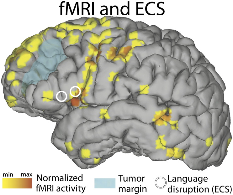
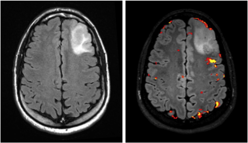
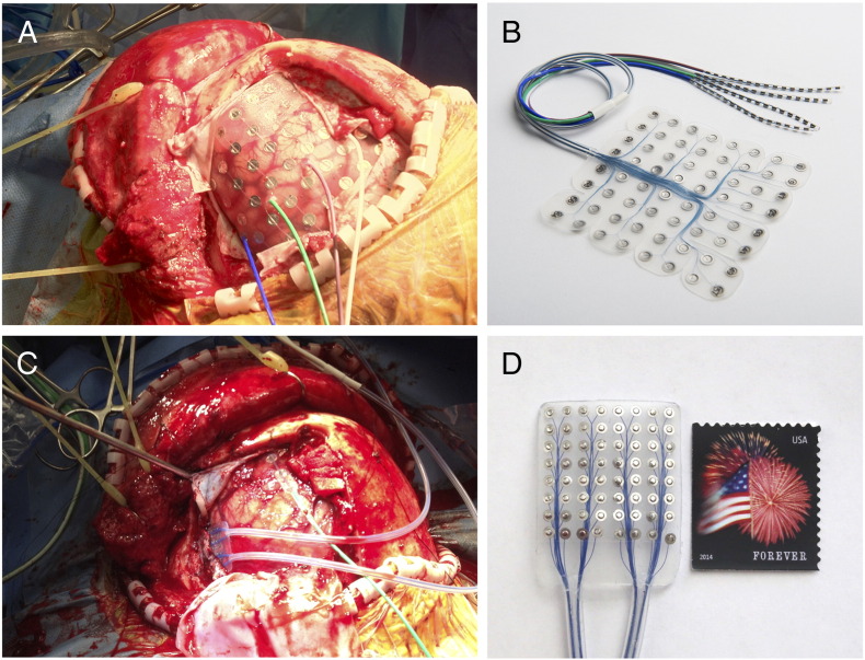
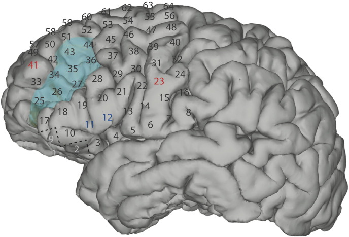
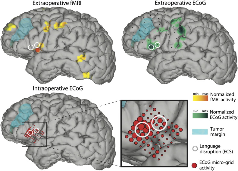
In this case report, we investigated the utility and practicality of passive intraoperative functional mapping of expressive language cortex using high-resolution electrocorticography (ECoG). The patient presented here experienced new-onset seizures caused by a medium-grade tumor in very close proximity to expressive language regions. In preparation of tumor resection, the patient underwent multiple functional language mapping procedures. We examined the relationship of results obtained with intraoperative high-resolution ECoG, extraoperative ECoG utilizing a conventional subdural grid, extraoperative electrical cortical stimulation (ECS) mapping, and functional magnetic resonance imaging (fMRI). Our results demonstrate that intraoperative mapping using high-resolution ECoG is feasible and, within minutes, produces results that are qualitatively concordant to those achieved by extraoperative mapping modalities. They also suggest that functional language mapping of expressive language areas with ECoG may prove useful in many intraoperative conditions given its time efficiency and safety. Finally, they demonstrate that integration of results from multiple functional mapping techniques, both intraoperative and extraoperative, may serve to improve the confidence in or precision of functional localization when pathology encroaches upon eloquent language cortex.
Keywords: Electrical cortical stimulation; Electrocorticography; High-density grid; Intraoperative; Language mapping; fMRI.
Figures
References
-
- Penfield W., Boldrey E. Somatic motor and sensory representation in the cerebral cortex of man as studied by electrical stimulation. Brain. 1937;60(4):389–443.
-
- Chakraborty A., McEvoy A.W. Presurgical functional mapping with functional MRI. Curr Opin Neurol. 2008;21(4):446–451. - PubMed
-
- Crone N.E., Miglioretti D.L., Gordon B., Sieracki J.M., Wilson M.T., Uematsu S. Functional mapping of human sensorimotor cortex with electrocorticographic spectral analysis. I. Alpha and beta event-related desynchronization. Brain. 1998;121(Pt 12):2271–2299. - PubMed
-
- Leuthardt E.C., Miller K., Anderson N.R., Schalk G., Dowling J., Miller J. Electrocorticographic frequency alteration mapping: a clinical technique for mapping the motor cortex. Neurosurgery. 2007;60(4 Suppl 2):260–270. [discussion 70-1] - PubMed
Grants and funding
LinkOut - more resources
Full Text Sources
Other Literature Sources
