

Clinical utility of indigenously formulated single-vial lyophilized HYNIC-TOC kit in evaluating Gastro-entero Pancreatic neuro endocrine tumours
- PMID: 27408857
- PMCID: PMC4937710
Clinical utility of indigenously formulated single-vial lyophilized HYNIC-TOC kit in evaluating Gastro-entero Pancreatic neuro endocrine tumours
Abstract
Objectives: The objective of this study was to evaluate the performance and utility of (99m)Tc HYNIC-TOC planar scintigraphy and SPECT/CT in the diagnosis, staging and management of gastroenteropancreatic neuroendocrine tumors (GPNETs).
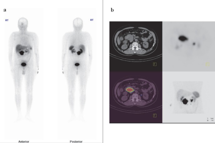
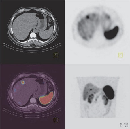
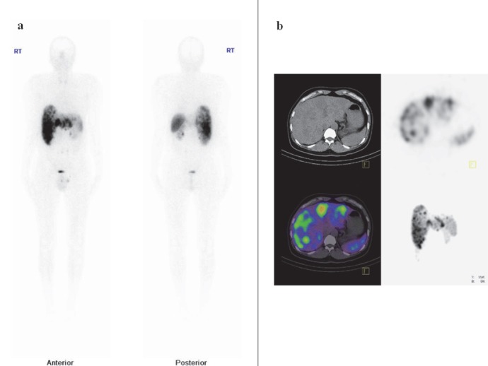
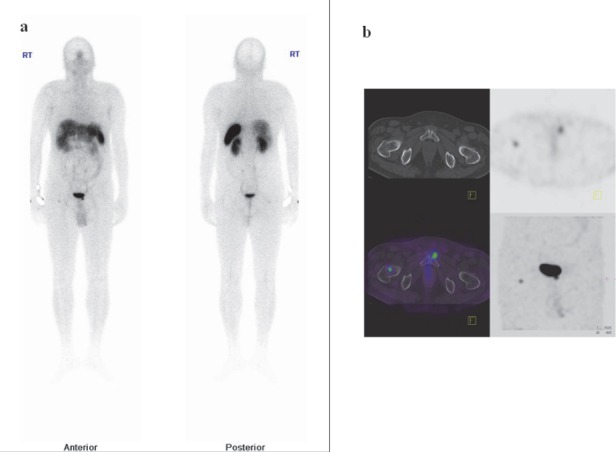
Methods: 22 patients (median age, 46 years) with histologically proven gastro- entero- pancreatic NETs underwent (99m)Tc HYNIC-TOC whole body scintigraphy and regional SPECT/CT as indicated. Scanning was performed after injection of 370-550 MBq (10-15 mCi) of (99m)Tc HYNIC-TOC intravenously. Images were evaluated by two experienced nuclear medicine physicians both qualitatively as well as semi quantitatively (tumor to background and tumor to normal liver ratios on SPECT -CT images). Results of SPECT/CT were compared with the results of conventional imaging. Histopathology results and follow-up somatostatin receptor scintigraphy with (99m)Tc HYNIC TOC or conventional imaging with biochemical markers were considered to be the reference standards.
Results: (99m)Tc HYNIC TOC showed sensitivity and specificity of 87.5% and 85.7%, respectively, for primary tumor and 100% and 86% for metastases. It was better than conventional imaging modalities for the detection of both primary tumor (P<0.001) and metastases (P<0.0001). It changed the management strategy in 6 patients (31.8%) and supported management decisions in 8 patients (36.3%).
Conclusion: (99m)Tc HYNIC TOC SPECT/CT appears to be a highly sensitive and specific modality for the detection and staging of GPNETs. It is better than conventional imaging for the evaluation of GPNETs and can have a significant impact on patient management and planning further therapeutic options.
Keywords: HYNIC TOC; Neuroendocrine tumor; SPECT CT.
Figures
References
-
- Modlin IM, Lye KD, Kidd M. A 5-decade analysis of 13,715 carcinoid tumors. Cancer. 2003;97:934–59. - PubMed
-
- Sutliff VE, Doppman JL, Gibril F, Venzon D J, Yu F, Serrano J, et al. Growth of newly diagnosed, untreated metastatic gastrinomas and predictors of growth patterns. J Clin Oncol. 1997;15:2420–31. - PubMed
-
- Fraker DL, Jensen RT. Pancreatic endocrine tumors. In: DeVita VT, Hellman S, Rosemberg SA, editors. Cancer: principles and practice of oncology. 5th ed. Philadelphia: PA: Lippincott-Raven; 1997. pp. 1678–1704.
-
- Eriksson B, Oberg K, Skogseid B. Neuroendocrine pancreatic endocrine tumors: clinical findings in a prospective study of 84 patients. Acta Oncol. 1989;28:373–7. - PubMed
-
- Schillaci O, Spanu A, Scopinaro F, Falchi A, Danieli R, Marongiu P, et al. Somatostatin receptor scintigraphy in liver metastasis detection from gastroenteropancreatic neuroendocrine tumors. J Nucl Med. 2003;44:359–68. - PubMed
LinkOut - more resources
Full Text Sources