

(18)F-FDG PET/CT in Neurolymphomatosis: Report of 3 Cases
- PMID: 27408859
- PMCID: PMC4937712
(18)F-FDG PET/CT in Neurolymphomatosis: Report of 3 Cases
Abstract
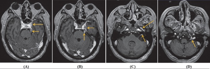
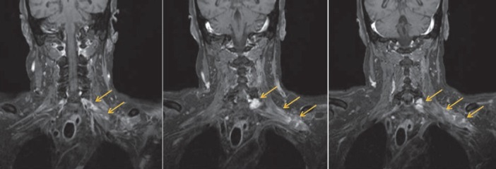
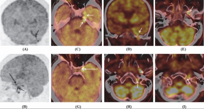
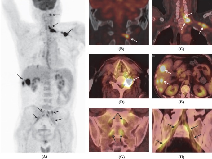
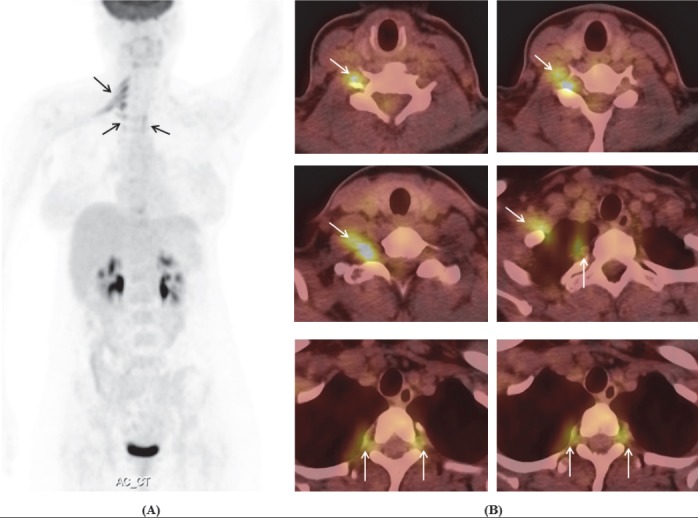
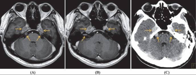
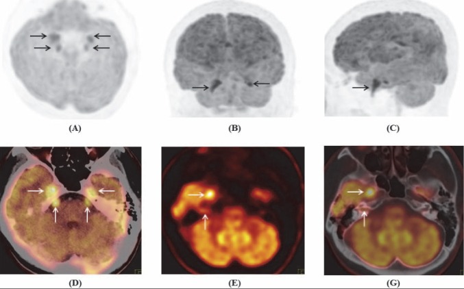
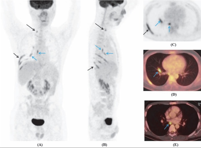
Neurolymphomatosis is a rare manifestation of non-Hodgkin lymphoma characterized by infiltration of peripheral nerves, nerve roots, plexus and cranial nerves by malignant lymphocytes. This report presents positron emission tomography/computed tomography (PET/CT)imaging with 2-deoxy-2-(18)F-fluoro-D-glucose ((18)F-FDG) in 3 cases of non-Hodgkin lymphoma with nerve infiltration, including one newly diagnosed lymphoma, one recurrent lymphoma in previous nerve lesions and one newly recurrent lymphoma. PET/CT could reveal the affected neural structures including cranial nerves, spinal nerve roots, brachial plexus, cervicothoracic ganglion, intercostal nerves, branches of the vagus nerve, lumbosacral plexus and sciatic nerves. There was relative concordance between PET/CT and MRI in detection of affected cranial nerves. PET/CT seemed to be better than MRI in detection of affected peripheral nerves. (18)F-FDG PET/CT was a whole-body imaging technique with the ability to reveal the affected cranial nerves, peripheral nerves, nerve roots and plexus in non-Hodgkin lymphoma. A thorough understanding of disease and use of advanced imaging modalities will increasingly detect neurolymphomatosis.
Keywords: 18F-FDG; Nerve; Neurolymphomatosis; PET/CT; Plexus.
Figures
Similar articles
-
18F-FDG PET/CT/MRI Fusion Images Showing Cranial and Peripheral Nerve Involvement in Neurolymphomatosis.Indian J Nucl Med. 2017 Jan-Mar;32(1):77-78. doi: 10.4103/0972-3919.198502. Indian J Nucl Med. 2017. PMID: 28242998 Free PMC article.
-
Neurolymphomatosis on F-18 FDG PET/CT and MRI Findings: A Case Report.Nucl Med Mol Imaging. 2011 Mar;45(1):76-8. doi: 10.1007/s13139-010-0070-8. Epub 2010 Dec 10. Nucl Med Mol Imaging. 2011. PMID: 24899982 Free PMC article.
-
Diagnostic utility of FDG-PET in neurolymphomatosis: report of five cases.J Neurol. 2016 Sep;263(9):1719-26. doi: 10.1007/s00415-016-8190-4. Epub 2016 Jun 10. J Neurol. 2016. PMID: 27286845
-
The role of 18F-FDG PET/CT in Neurolymphomatosis: A Comprehensive Imaging Approach.J Pak Med Assoc. 2024 Apr;74(4):822-824. doi: 10.47391/JPMA.24-30. J Pak Med Assoc. 2024. PMID: 38751291 Review.
-
Recurrence of nasal type NK/T cell lymphoma presenting as neurolymphomatosis on 18F-FDG PET/CT: A case report and literature review.Medicine (Baltimore). 2020 Jan;99(1):e18640. doi: 10.1097/MD.0000000000018640. Medicine (Baltimore). 2020. PMID: 31895825 Free PMC article. Review.
Cited by
-
Neurolymphomatosis: a single-center experience of neuromuscular manifestations, treatments, and outcomes.J Neurol. 2021 Mar;268(3):851-859. doi: 10.1007/s00415-020-10202-0. Epub 2020 Oct 23. J Neurol. 2021. PMID: 33098033
-
B-cell peripheral neurolymphomatosis: MRI and 18F-FDG PET/CT imaging characteristics.Skeletal Radiol. 2019 Jul;48(7):1043-1050. doi: 10.1007/s00256-019-3145-3. Epub 2019 Jan 22. Skeletal Radiol. 2019. PMID: 30666391
-
Neurolymphomatosis in Recrudescent Diffuse Large B-cell Lymphoma.Asia Ocean J Nucl Med Biol. 2023;11(1):89-92. doi: 10.22038/AOJNMB.2022.66666.1464. Asia Ocean J Nucl Med Biol. 2023. PMID: 36619186 Free PMC article.
-
Diagnostic delay in a case of T-cell neurolymphomatosis.BMJ Case Rep. 2019 Dec 29;12(12):e232538. doi: 10.1136/bcr-2019-232538. BMJ Case Rep. 2019. PMID: 31888900 Free PMC article.
-
The natural history of neurolymphomatosis.BJC Rep. 2024 Apr 23;2(1):34. doi: 10.1038/s44276-024-00053-x. BJC Rep. 2024. PMID: 39516680 Free PMC article.
References
-
- Anh PT, Duc NB. The situation with cancer control in Vietnam. Jpn J Clin Oncol. 2002;32(Suppl):S92–7. - PubMed
-
- Baehring JM, Batchelor TT. Diagnosis and management of neurolymphomatosis. Cancer J. 2012;18(5):463–8. - PubMed
-
- Allen-Auerbach M, de Vos S, Czernin J. PET/computed tomography and lymphoma. Radiol Clin North Am. 2013;51(5):833–44. - PubMed
Publication types
LinkOut - more resources
Full Text Sources