. 2016 Jul 13;12(7):e1005710.
doi: 10.1371/journal.ppat.1005710. eCollection 2016 Jul.

The Actin Filament-Binding Protein Coronin Regulates Motility in Plasmodium Sporozoites

Affiliations

The Actin Filament-Binding Protein Coronin Regulates Motility in Plasmodium Sporozoites

Kartik S Bane et al. PLoS Pathog. .

Erratum in

Abstract

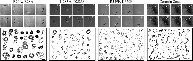
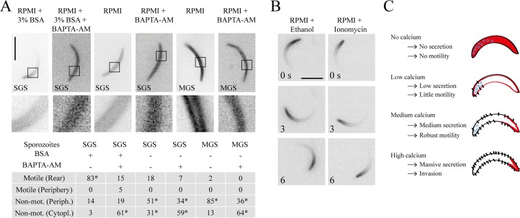
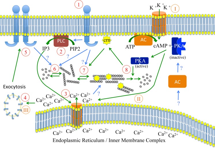
Parasites causing malaria need to migrate in order to penetrate tissue barriers and enter host cells. Here we show that the actin filament-binding protein coronin regulates gliding motility in Plasmodium berghei sporozoites, the highly motile forms of a rodent malaria-causing parasite transmitted by mosquitoes. Parasites lacking coronin show motility defects that impair colonization of the mosquito salivary glands but not migration in the skin, yet result in decreased transmission efficiency. In non-motile sporozoites low calcium concentrations mediate actin-independent coronin localization to the periphery. Engagement of extracellular ligands triggers an intracellular calcium release followed by the actin-dependent relocalization of coronin to the rear and initiation of motility. Mutational analysis and imaging suggest that coronin organizes actin filaments for productive motility. Using coronin-mCherry as a marker for the presence of actin filaments we found that protein kinase A contributes to actin filament disassembly. We finally speculate that calcium and cAMP-mediated signaling regulate a switch from rapid parasite motility to host cell invasion by differentially influencing actin dynamics.


Conflict of interest statement

I have read the journal's policy and the authors of this manuscript have the following competing interests: KH is shareholder of MalVa GmbH, a spin-off company of the Heidelberg University Hospital focussing on malaria vaccine development. There are no patents, products in development, marketed products or financial interests to declare. This does not alter our adherence to all PLOS Pathogens policies on sharing data and materials.

Figures

Fig 1
Fig 1. Coronin is important for directional motility and localizes to the rear in an actin-dependent manner.
(A) Wild type (WT) P. berghei strain ANKA parasites (i) and coronin(-) (ii-iv) sporozoites were allowed to settle on a glass-bottom Petri-dish and imaged with differential interference contrast microscopy. WT sporozoites move in near-perfect circles with an average speed of about 2 μm/s, while coronin(-) sporozoites frequently fail to move or undergo different types of non-persistent movements such as sporadic translocation [37] or various types of flexing behavior (iii-iv). Scale bar: 10 μm. (B) Sporozoites as imaged in A were tracked using ImageJ. Tracks of randomly selected WT sporozoites reveal the persistent circular movement while tracks of coronin(-) sporozoites reveal non-circular trajectories. Scale bar: 10 μm. (C) Parasites expressing coronin-mCherry were generated (see S4A Fig) and sporozoites imaged in RPMI medium with a fluorescence microscope in the presence and absence of the motility stimulating bovine serum albumin (BSA). Non-motile sporozoites isolated from the mosquito midgut (MGS) or salivary gland (SGS) showed peripheral fluorescence. Motile salivary gland derived sporozoites showed a fluorescent signal at the rear end. Note the high background in the non-motile sporozoite images indicating the low expression level of coronin-mCherry. There is no quantitative difference in expression level of activated and non-activated sporozoites. Scale bar: 5 μm. (D) Motile sporozoites expressing coronin-mCherry were imaged with a fluorescence microscope under various concentrations of the actin dynamics modulating compounds cytochalasin D and jasplakinolide as indicated at the top right of each panel (in nM). Note the relocalization of the fluorescence signal with increasing drug concentrations. Bars in the top left panel indicate lines where fluorescent intensity profiles were taken to determine front versus rear (F/R) intensity ratios of coronin-mCherry. A low F/R ratio indicates localization to the rear end. Scale bar: 5 μm. (E) Quantitative assessment of the F/R ratio from at least 10 images taken under the indicated concentrations.
Fig 2
Fig 2. Mutant coronins reveal distinct binding to membranes and actin filaments.
(A) Multiple sequence alignment shows that amino acids found to be important for actin binding in yeast are conserved between mouse, yeast and apicomplexans and are marked in red. (B) Fluorescence microscopy of a parasite line overexpressing coronin-mCherry from the uis3 promoter. Note the peripheral fluorescence in non-motile (RPMI) and motile (RPMI+BSA) salivary gland derived sporozoites. Numbers indicate time in seconds. Scale bar: 5 μm. (C) Localization of a number of overexpressed mutant coronin-mCherry fusion proteins. Note the three different types of localizations: cytoplasmic (coronin-8mut; K283A/E, D285A/R), peripheral (R24E, R28E; R349A/E, K350A/E) and polarized (WT, R24A, R28A). Scale bar: 5 μm. (D) Graph showing a quantitative assessment of the front versus rear ratio from 20 images of the various parasite lines overexpressing mutated coronin-mCherry as indicated on the x-axis. (E) The peripheral localization of R349A/E, K350A/E mutants is not altered when sporozoites are incubated with cytochalasin (100 nM), jasplakinolide (100 nM), ionomycin (1000 nM) or cytochalasin D (100 nM) + ionomycin (500 nM) Scale bar: 5 μm.
Fig 3
Fig 3. Mutant coronins exhibit defects in sporozoite motility.
Time-lapse images and randomly selected tracks of different parasite lines expressing endogenous coronin fused to mCherry with the indicated mutations in their putative actin-binding sites. Note that R24A, R28A moves in a similar manner to WT, while the other mutants move differently and exhibit bending movements so far not seen in any wild type or mutated sporozoite, most strikingly seen in the time lapse of a sporozoites expressing the R349E, K350E mutation. A small but discernable fraction of sporozoite expressing the K283A, D285A mutation in coronin can move in persistent circles. Scale bars: 5 μm (images), 10 μm (tracks).
Fig 4
Fig 4. Intracellular calcium release precedes the relocalization of coronin and motility.
(A) Activated and non-activated sporozoites overexpressing coronin-mCherry were treated with or without BAPTA-AM to investigate effects on motility and coronin-mCherry localization with fluorescence microscopy. Note that BAPTA-AM ceases motility and relocalizes coronin from the periphery to the cytoplasm in activated salivary gland sporozoites (panel 1–2, top; p < 0.0001). Treatment with BAPTA-AM of non-activated salivary gland sporozoites (panel 3–4, top; p < 0.0002) and midgut sporozoites (panel 5–6, top; p < 0.0001) relocalizes coronin to the cytoplasm. Scale bar: 5 μm. The table shows the percentage of motile and non-motile sporozoites as determined from imaging at least 100 sporozoites and their associated localization pattern of coronin-mCherry [bottom]. The statistical differences are calculated by Fisher’s exact test. (B) Addition of ethanol or of the ionophore ionomycin to non-activated sporozoites leads to coronin-mCherry relocalization to the rear and motility. Scale bar: 5 μm. (C) Model of the role of calcium mediated receptor secretion on sporozoite progression from mosquito to the liver. In the absence of cytosolic calcium coronin (red) localizes to the sporozoite cytoplasm. At low calcium concentrations coronin localizes to the periphery and few TRAP family adhesins are released onto the sporozoite surface. At medium calcium concentrations, coronin localizes to the rear and more adhesins are released onto the sporozoite surface. Signaling associated with these events leads to optimal motility. Finally at very high calcium concentrations massive secretion leads a further increase of adhesins on the surface, which leads to invasion [49].
Fig 5
Fig 5. cAMP signaling downstream of coronin relocalization modulates motility.
(A) Sporozoites were incubated in RPMI + 3% BSA with the indicated kinase inhibitors at the indicated concentrations, imaged and their movement pattern quantified as gliding, waving or attached. The inhibitors stopped sporozoite motility in a dose-dependent fashion. Over 150 sporozoites were examined for each experiment. Significant differences (Fisher’s exact test) from the controls show both concentrations of H89 tested (p<0.0001) as well as 500 μM SQ22536 (SQ) (p = 0.01; p = 0.46 for 200 μM SQ22536). (B) Forskolin partially stimulates sporozoite motility when added to sporozoites in RPMI. Over 300 sporozoites were examined and their motility pattern quantified as in A. The shown difference is significant (p<0.0001; Fisher’s exact test). (C) Coronin-mCherry expressing sporozoites were investigated under various kinase inhibitors (each at 0,1 mM) with a fluorescence microscope to determine coronin-mCherry localization. The fluorescence stays at the rear in activated sporozoites arrested with PKA inhibitors. Coronin-mCherry relocalizes to the periphery when additional cytochalasin D is applied. Addition of forskolin to non-activated sporozoites leads to the localization of coronin-mCherry to the rear. Scale bar: 5 μm. (D) Table showing percentages of motile and non-motile sporozoites and the associated localization patterns (rear versus non-rear) of coronin-mCherry under the indicated conditions; low and high concentrations of SQ22536 (SQ) were 0,1 and 0,5 mM, respectively. Between 105 and 120 sporozoites were examined per condition. Statistical differences determined by Fisher’s exact test was p<0.0001 from the respective controls listed in the table of Fig 4A. (E) Examples of FRAP of motile sporozoites with coronin-mCherry localized to the rear. Scale bar: 5 μm. Circle indicates location of the bleach spot. (F) Quantitative analysis of FRAP. Coronin-mCherry recovers as fast in motile (>0.25 μm/s) as in non-motile (<0.25 μm/s) sporozoites if pooled across all conditions. There is also no difference in recovery time depending on the localization of coronin-mCherry [top graphs]. Quantitative analysis of FRAP data over a range of conditions [bottom graph]. Average values (+/- S.D.) are indicated above the graph. Bars show significant differences (* p<0.05; ** p<0.01), non-significance (ns) or p-values (Students t-test). Coronin-mCherry recovers significantly faster in non-motile sporozoites incubated in RPMI than in any other condition. With all other conditions there is no difference from each other with the exception of H89, where coronin-mCherry recovers significantly slower when compared to controls, 1 μM Cytochalasin D and 100 nM Jasplakinolide [bottom].
Fig 6
Fig 6. Speculative working model on coronin function at the interface between calcium and cAMP signaling during motility (red Arab numbers) and invasion (orange Roman numbers).
Upon activation of a trans-membrane receptor by extracellular ligands on the salivary gland or in the skin (1), phospholipase C is activated and converts PIP2 into IP3 (2). This leads to the release of calcium from intracellular stores (3) and subsequent exocytosis of micronemes, which bring more receptors to the plasma membrane (5). These reinforce signaling and activate actin polymerization (6). Actin filaments are organized by surface receptors and relocalization of coronin from peripheral membranes (either PM or IMC) to actin filaments (7). This leads to efficient adhesion and force production essential for motility in 2D. Coronin (crn) relocalizes from actin filaments as these are disassembled through the action of PKA, which is possibly activated by cytosolic adenylate cyclase (AC). Upon stimulation of membrane bound adenylate cyclase more cAMP is produced leading to higher PKA activity and further calcium release from intracellular stores. The additionally released receptors then mediate invasion.

Similar articles

Cited by

References

    1. Douglas RG, Amino R, Sinnis P, Frischknecht F (2015) Active migration and passive transport of malaria parasites. Trends Parasitol 31: 357–362. 10.1016/j.pt.2015.04.010 - DOI - PubMed
    1. Vanderberg JP, Frevert U (2004) Intravital microscopy demonstrating antibody-mediated immobilisation of Plasmodium berghei sporozoites injected into skin by mosquitoes. Int J Parasitol 34: 991–996. - PubMed
    1. Amino R, Thiberge S, Martin B, Celli S, Shorte S, et al. (2006) Quantitative imaging of Plasmodium transmission from mosquito to mammal. Nat Med 12: 220–224. - PubMed
    1. Menard R, Tavares J, Cockburn I, Markus M, Zavala F, et al. (2013) Looking under the skin: the first steps in malarial infection and immunity. Nat Rev Microbiol 11: 701–712. 10.1038/nrmicro3111 - DOI - PubMed
    1. Prudencio M, Rodriguez A, Mota MM (2006) The silent path to thousands of merozoites: the Plasmodium liver stage. Nat Rev Microbiol 4: 849–856. - PubMed

Publication types