MiR-27a-3p functions as an oncogene in gastric cancer by targeting BTG2
- PMID: 27409164
- PMCID: PMC5239526
- DOI: 10.18632/oncotarget.10460
MiR-27a-3p functions as an oncogene in gastric cancer by targeting BTG2
Abstract
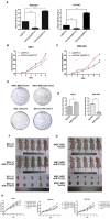
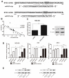
microRNA-27a (miR-27a) is frequently dysregulated in human carcinoma, including gastric cancer. The B-cell translocation gene 2 (BTG2) has been implicated in gastric carcinogenesis. However, till now, the link between miR-27a and BTG2 in gastric cancer has not been reported. Here, we found that two isoforms of mature miR-27a, miR-27a-5p and miR-27-3p, were both frequently overexpressed in gastric cancer tissues and cell lines, whereas the expression level of miR-27-3p in gastric cancer was significantly higher than that of miR-27a-5p. And overexpression of miR-27a-3p, but not miR-27a-5p, markedly promoted gastric cancer cell proliferation in vitro as well as tumor growth in vivo. Further experiments revealed that BTG2 was a direct and functional target of miR-27a-3p in gastric cancer and miR-27a-3p inhibition obviously up-regulated the expression of BTG2. In turn, overexpression of BTG2 triggered G1/S cell cycle arrest, induced subsequent apoptosis, and inhibited C-myc activation following Ras/MEK/ERK signaling pathway, which involved in the biological effects of miR-27a-3p/BTG2 axis on gastric carcinogenesis and cancer progression. Overall, these results suggested that the miR-27a-3p/BTG2 axis might represent a promising diagnostic biomarker for gastric cancer patients and could be a potential therapeutic target in the management of gastric cancer.
Keywords: BTG2; apoptosis; cell proliferation; gastric cancer; miR-27a-3p.
Conflict of interest statement
The authors have no competing financial or intellectual interests.
Figures


Similar articles
-
MiR-25-3p promotes the proliferation of triple negative breast cancer by targeting BTG2.Mol Cancer. 2018 Jan 8;17(1):4. doi: 10.1186/s12943-017-0754-0. Mol Cancer. 2018. PMID: 29310680 Free PMC article.
-
MiR-6875-3p promotes the proliferation, invasion and metastasis of hepatocellular carcinoma via BTG2/FAK/Akt pathway.J Exp Clin Cancer Res. 2019 Jan 8;38(1):7. doi: 10.1186/s13046-018-1020-z. J Exp Clin Cancer Res. 2019. PMID: 30621734 Free PMC article.
-
Extracellular vesicles carry miR-27a-3p to promote drug resistance of glioblastoma to temozolomide by targeting BTG2.Cancer Chemother Pharmacol. 2022 Feb;89(2):217-229. doi: 10.1007/s00280-021-04392-1. Epub 2022 Jan 17. Cancer Chemother Pharmacol. 2022. PMID: 35039898
-
BTG2: a rising star of tumor suppressors (review).Int J Oncol. 2015 Feb;46(2):459-64. doi: 10.3892/ijo.2014.2765. Epub 2014 Nov 18. Int J Oncol. 2015. PMID: 25405282 Review.
-
Significance of microRNA 21 in gastric cancer.Clin Res Hepatol Gastroenterol. 2016 Nov;40(5):538-545. doi: 10.1016/j.clinre.2016.02.010. Epub 2016 May 11. Clin Res Hepatol Gastroenterol. 2016. PMID: 27179559 Review.
Cited by
-
MiR-27a-3p promotes the osteogenic differentiation by activating CRY2/ERK1/2 axis.Mol Med. 2021 Apr 26;27(1):43. doi: 10.1186/s10020-021-00303-5. Mol Med. 2021. PMID: 33902432 Free PMC article.
-
Oncogenic long intervening noncoding RNA Linc00284 promotes c-Met expression by sponging miR-27a in colorectal cancer.Oncogene. 2021 Jun;40(24):4151-4166. doi: 10.1038/s41388-021-01839-w. Epub 2021 May 28. Oncogene. 2021. PMID: 34050266 Free PMC article.
-
MicroRNA-539 inhibits colorectal cancer progression by directly targeting SOX4.Oncol Lett. 2018 Aug;16(2):2693-2700. doi: 10.3892/ol.2018.8892. Epub 2018 Jun 4. Oncol Lett. 2018. PMID: 30013665 Free PMC article.
-
Effects of tRNA-derived fragments and microRNAs regulatory network on pancreatic acinar intracellular trypsinogen activation.Bioengineered. 2022 Feb;13(2):3207-3220. doi: 10.1080/21655979.2021.2018880. Bioengineered. 2022. PMID: 35045793 Free PMC article.
-
MiR-25-3p promotes the proliferation of triple negative breast cancer by targeting BTG2.Mol Cancer. 2018 Jan 8;17(1):4. doi: 10.1186/s12943-017-0754-0. Mol Cancer. 2018. PMID: 29310680 Free PMC article.
References
-
- Jemal A, Siegel R, Xu J, Ward E. Cancer statistics, 2010. CA Cancer J Clin. 2010;60:277–300. - PubMed
-
- Jemal A, Bray F, Center MM, Ferlay J, Ward E, Forman D. Global cancer statistics. CA Cancer J Clin. 2011;61:69–90. - PubMed
-
- Liu T, Tang H, Lang Y, Liu M, Li X. MicroRNA-27a functions as an oncogene in gastric adenocarcinoma by targeting prohibitin. Cancer letters. 2009;273:233–242. - PubMed
-
- Yang Q, Jie Z, Ye S, Li Z, Han Z, Wu J, Yang C, Jiang Y. Genetic variations in miR-27a gene decrease mature miR-27a level and reduce gastric cancer susceptibility. Oncogene. 2012;33:193–202. - PubMed
MeSH terms
Substances
LinkOut - more resources
Full Text Sources
Other Literature Sources
Medical
Miscellaneous

