. 2016 Aug 2;7(31):50564-50574.
doi: 10.18632/oncotarget.10501.

TREM-1 expression in craniopharyngioma and Rathke's cleft cyst: its possible implication for controversial pathology

Affiliations

TREM-1 expression in craniopharyngioma and Rathke's cleft cyst: its possible implication for controversial pathology

Yi Liu et al. Oncotarget. .

Abstract

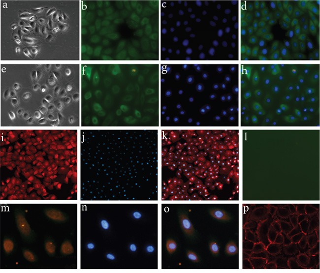
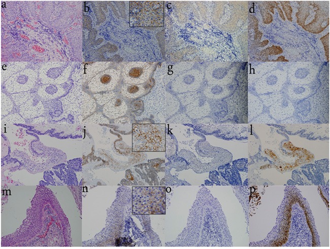
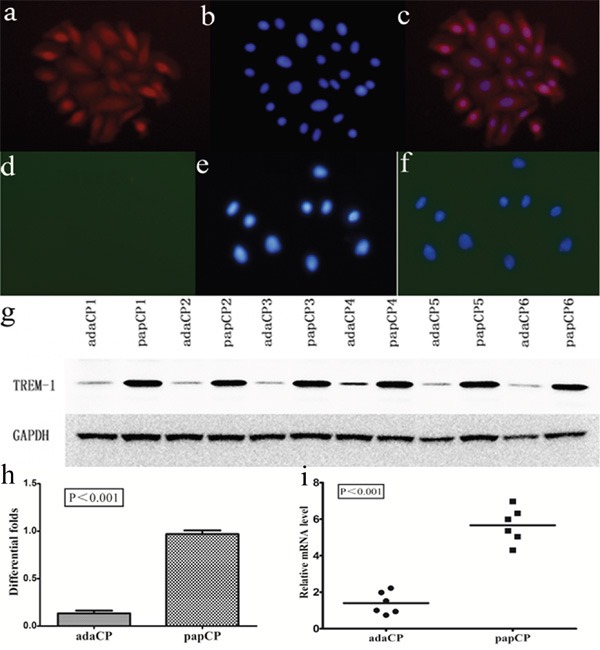
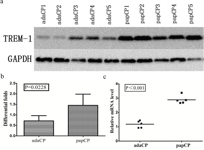
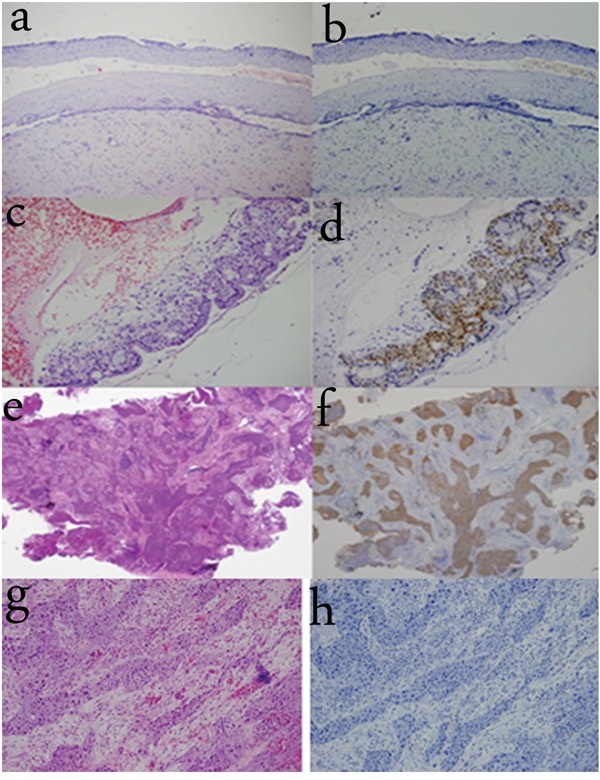
Whether a mixed type of craniopharyngioma (CP) exists and whether papillary craniopharyngioma (pCP) is on a histopathological continuum with Rathke's cleft cyst (RCC) remain controversial. Herein, we examined the expression and localization of β-catenin, BRAF p.V600E (V600E), and triggering receptor expressed on myeloid cells-1 (TREM-1) in 58 samples including 20 pCPs, 26 adamantinomatous craniopharyngiomas (aCP), and 12 RCCs. Five aCPs were diagnosed with mixed type CPs and the remaining 21 cases were pure aCPs. Four of the 12 RCCs presented with significant squamous epithelium (SE). V600E immunoreactivity was observed in all pCPs in the cytoplasm, but not in the nuclei. aCPs and RCCs, including mixed type CP, did not express V600E. Nuclear β-catenin translocation was detected exclusively in aCPs. TREM-1 was expressed in pCPs. Additionally, TREM-1 expression was detected in the SE of 5 "mixed type" CPs, while it was absent in pure aCPs. TREM-1 was expressed in 4 RCCs with SE, but not in the remaining 8 RCCs. TREM-1 mRNA levels were compared in cultured pCP and aCP cells. TREM-1 mRNA level was significantly (p < 0.001; up to 4.045 fold) higher in pCPs than in aCPs. Western blotting revealed a significantly (p < 0.001; up to 7.19 fold) lower level of TREM-1 expression in aCP cells compared to that in pCP cells. Our findings further supported that RCC and pCP may represent two ends of a morphological spectrum. A variant showing overlapping histological features of aCP and pCP should not be considered as a mixed type.

Keywords: Rathke’s cleft cyst; SE; TREM-1; craniopharyngioma; metaplasia.


Conflict of interest statement

The authors declare that they have no conflicts of interest.

Figures

Figure 1
Figure 1. Primary cell culture of CP samples
a-d. Primary cell culture of aCP; a. morphology of aCP cells; b-d. aCP cells are positive for pan-CK. (400×) e-h. Primary cell culture of pCP; e. morphology of pCP cells; f-h. pCP cells are positive for pan-CK.(400×) i-l. β-catenin and V600E expression in cultured aCP cells; i-k. nuclear β-catenin translocation is detected in cultured aCP cells; l. V600E immunoreactivity is absent in cultured aCP cells. (400×) m-p. β-catenin and V600E expression in cultured pCP cells; m-o. V600E immunoreactivity is observed in cultured pCP cells with strong detection in the cytoplasm; p. β-catenin is restricted to the cell membrane of cultured pCP cells. (400×)
Figure 2
Figure 2. Differential distribution pattern of β-catenin, V600E, and TREM-1 in CPs and RCCs
a. H&E staining of pCP indicates the squamous and well-differentiated, non-keratinizing epithelium. (200×) b. Nuclear β-catenin translocation is not observed and β-catenin expression is restricted to the cell membrane. (200×) c. V600E immunoreactivity is observed in the SE of pCPs with strong detection in the cytoplasm, while it is not detected in the nuclei. (200×) d. TREM-1 immunoreactivity is strongly detected in the cytoplasm and nuclei of the SE in all pCPs. (200×) e. H&E staining of pure aCP shows the classical appearance with peripheral palisading epithelium, loose ‘stellate reticulum’, and whorl-like cells. (200×) f. β-catenin translocation is observed especially in whorl-like cells. (200×) g. V600E immunoreactivity is not detected in pure aCP tissues. (200×) h. TREM-1 immunoreactivity is also absent in pure aCP tissues. (200×) i. H&E staining of “mixed type CPs” shows both SE and peripheral palisading epithelium, whorl-like cells are also detected. (200×) j. β-catenin translocation is observed in whorl-like cells and is restricted to the cell membrane in SE of “mixed type CP”. (200×) k. V600E immunoreactivity is absent in SE and other components of “mixed type CP”. (200×) l. TREM-1 immunoreactivity is observed in the SE of “mixed type CP” and absent in other components of “mixed type CPs”. (200×) m. H&E staining of RCC with stratified SE. (400×) n. No nuclear β-catenin translocation is observed and β-catenin expression is restricted to the cell membrane. (400×) o. V600E immunoreactivity is absent in SE. (400×) p. TREM-1 immunoreactivity is observed in the SE. (400×)
Figure 3
Figure 3. TREM-1 expression pattern in aCP and pCP cells
a-c. TREM-1 expression is strongly detected in the cytoplasm and nuclei of pCP cells. (400×) d-f. TREM-1 expression is absent in aCP cells. (400×) g-h. Western blotting shows the protein level of TREM-1 in cultured CP cells. Evaluation of the relative TREM-1 expression revealed a significantly (p < 0.001; up to 7.19 fold) lower level in aCP (mean = 0.13 ± 0.07) compared to that of the pCP variant (mean = 0.97 ± 0.03) i. TREM-1 mRNA levels of aCP and pCP cells were compared by qRT-PCR. TREM-1 relative expression level was significantly (p < 0.001; up to 4.045 fold) higher in pCP (mean = 5.66 ± 0.39) compared to that of the aCP variant (mean = 1.40 ± 0.25).
Figure 4
Figure 4. TREM-1 expression pattern in aCP and pCP tissues
a-b. Western blotting shows the protein level of TREM-1 in CP tissues. TREM-1 expression level is significantly (p = 0.0228; up to 1.89 fold) lower in aCP tissues (mean = 0.714 ±0.112) compared to that of the pCP variant (mean = 1.453 ± 0.238). c. Evaluation of the relative TREM-1 expression revealed a significantly (p < 0.001; up to 2.08 fold) higher level in pCP (mean = 2.99 ± 0.12) compared to that of the aCP tissues (mean = 1.28 ± 0.11)
Figure 5
Figure 5. TREM-1 expression in other SE tissues
a-b. TREM-1 immunoreactivity is absent in normal cervical squamous epithelium. (100×) c-d. TREM-1 immunoreactivity is observed in metaplastic cervical squamous epithelium and strongly detected in the cytoplasm and nuclei. (200×) e-f. TREM-1 immunoreactivity is observed in squamous cell lung cancer (originating from metaplastic SE). (40×) g-h. TREM-1 immunoreactivity is absent in tongue carcinoma (originating from SE). (200×)

Similar articles

Cited by

References

    1. Larkin SJ, Ansorge O. Pathology and pathogenesis of craniopharyngiomas. Pituitary. 2013;16:9–17. - PubMed
    1. Martinez-Barbera JP, Buslei R. Adamantinomatous craniopharyngioma: pathology, molecular genetics and mouse models. J Pediatr Endocrinol Metab. 2015;28:7–17. - PubMed
    1. Prabhu VC, Brown HG. The pathogenesis of craniopharyngiomas. Childs Nerv Syst. 2005;21:622–627. - PubMed
    1. Zada G, Lin N, Ojerholm E, Ramkissoon S, Laws ER. Craniopharyngioma and other cystic epithelial lesions of the sellar region: a review of clinical, imaging, and histopathological relationships. Neurosurg Focus. 2010;28:E4. - PubMed
    1. Okada T, Fujitsu K, Ichikawa T, Mukaihara S, Miyahara K, Kaku S, Uryuu Y, Niino H, Yagishita S, Shiina T. Coexistence of adamantinomatous and squamous-papillary type craniopharyngioma: case report and discussion of etiology and pathology. Neuropathology. 2012;32:171–173. - PubMed

MeSH terms