. 2016 Aug 16;88(16):8264-71.
doi: 10.1021/acs.analchem.6b02104. Epub 2016 Jul 27.

Continuous On-Chip Cell Separation Based on Conductivity-Induced Dielectrophoresis with 3D Self-Assembled Ionic Liquid Electrodes

Affiliations

Continuous On-Chip Cell Separation Based on Conductivity-Induced Dielectrophoresis with 3D Self-Assembled Ionic Liquid Electrodes

Mingrui Sun et al. Anal Chem. .

Abstract

Dielectrophoresis (DEP) has been widely explored to separate cells for various applications. However, existing DEP devices are limited by the high cost associated with the use of noble metal electrodes, the need of high-voltage electric field, and/or discontinuous separation (particularly for devices without metal electrodes). We developed a DEP device with liquid electrodes, which can be used to continuously separate different types of cells or particles based on positive DEP. The device is made of polydimethylsiloxane (PDMS), and ionic liquid is used to form the liquid electrodes, which has the advantages of low cost and easy fabrication. Moreover, the conductivity gradient is utilized to achieve the DEP-based on-chip cell separation. The device was used to separate polystyrene microbeads and PC-3 human prostate cancer cells with 94.7 and 1.2% of the cells and microbeads being deflected, respectively. This device is also capable of separating live and dead PC-3 cancer cells with 89.8 and 13.2% of the live and dead cells being deflected, respectively. Moreover, MDA-MB-231 human breast cancer cells could be separated from human adipose-derived stem cells (ADSCs) using this device with high purity (81.8 and 82.5% for the ADSCs and MDA-MB-231 cells, respectively). Our data suggest the great potential of cell separation based on conductivity-induced DEP using affordable microfluidic devices with easy operation.


Figures
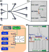
Figure 1
Figure 1
The DEP device with ionic liquid electrodes. (a) A sketch of the microfluidic channel system in the device. (b) A schematic of the experimental setup. (c) A zoom-in view of the ionic liquid electrodes and the main channel on the real image showing the stable interface formed between the ionic liquid and the DEP buffer. Scale bar: 100 μm. (d) A schematic showing the mechanism of the particle and cell separation. Particles 1 and 2 with different electrical properties and size are deflected by the DEP force differently and can be separated from each other in the electrode region. The dashed circles represent the intermediate position of the particles.
Figure 2
Figure 2
Analysis of conductivity and electric field distribution. (a) The simulated conductivity distribution in the first and second electrode regions. The conductivity of the modified DEP buffer is 14 mS/m, and the conductivity of the original DEP buffer is 1 mS/m. (b) The simulated electric field in the first and second electrode regions. The modified DEP buffer is introduced into I1 and the original DEP buffer is introduced into I2, when 88 V is applied on the device. (c) The distribution of Rhodamine B at the first and second electrodes regions without electric field. Scale bar: 100 μm. The velocity at the beginning of the main channel was set as 6 mm/s. The flow rate of the modified DEP buffer with Rhodamine B (10 μg/mL): 50 μL/hr, the flow rate of the original DEP buffer: 250 μL/hr.
Figure 3
Figure 3
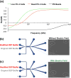
Mechanism analysis of cell deflection. (a) The real parts of the CM factors of live PC-3 cells, dead PC-3 cells, and 20 μm Polystyrene beads calculated with the two-layer model. The conductivity of the surrounding medium is set as 14 mS/m and its relative permittivity is set as 80. The conductivity of cell membrane and cell cytoplasm, the relative permittivity of cell membrane and cell cytoplasm of live PC-3 cells are set as 1×10⁻⁴ mS/m, 1000 mS/m, 20, and 60, respectively. The conductivity and relative permittivity of the polystyrene beads are set as 0.05 mS/m and 60, respectively. The conductivity of cell membrane and cell cytoplasm, the relative permittivity of cell membrane and cell cytoplasm of dead PC-3 cells are set as 1×10⁻² mS/m, 500 mS/m, 10, and 60, respectively. The diameter of the live and dead PC-3 cells is 16 μm. (b) The cells move out of the device without deflection, when modified DEP buffer is introduced from I1 (50 μL/hr) and original DEP buffer is introduced from I2 (250 μL/hr), and no electric field is applied. (c) The cells move out of the device with deflection, when modified DEP buffer is introduced from I1 (50 μL/hr) and original DEP buffer is introduced from I2 (250 μL/hr), and electric field is applied. Scale bar: 100 μm.
Figure 4
Figure 4
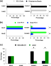
The separation of PC-3 cells and 20 μm polystyrene beads, live and dead PC-3 cells, and ADSCs and MDA-MB-231 cells. (a) The separation efficiency of PC-3 cells and 20 μm polystyrene beads when the voltage is 88 Vrms and the frequency is 100 kHz. (b) The separation efficiency of live and dead PC-3 cells when the voltage is 53 Vrms and the frequency is 50 kHz. (c) Purity of ADSCs and MDA-MB-231 cells in the outlet when the initial cell number ratio is 1:1. The voltage applied is 35 Vrms and the frequency is 50 kHz. Ncell > 500. Flow rates: 50 and 250 μL/hr for the modified and original DEP buffers, respectively.

Similar articles

Cited by

References

    1. Armstrong DW, Schneiderheinze JM, Kullman JP, He LF. Fems Microbiol Lett. 2001;194:33–37. - PubMed
    1. Cabrera CR, Yager P. Electrophoresis. 2001;22:355–362. - PubMed
    1. Fizazi K, Morat L, Chauveinc L, Prapotnich D, De Crevoisier R, Escudier B, Cathelineau X, Rozet F, Vallancien G, Sabatier L, Soria JC. Ann Oncol. 2007;18:518–521. - PubMed
    1. Girod M, Armstrong DW. Electrophoresis. 2002;23:2048–2056. - PubMed
    1. Hayes DF, Cristofanilli M, Budd GT, Ellis MJ, Stopeck A, Miller MC, Matera J, Allard WJ, Doyle GV, Terstappen LWWM. Clin Cancer Res. 2006;12:4218–4224. - PubMed

Publication types

LinkOut - more resources